To install click the Add extension button. That's it.
The source code for the WIKI 2 extension is being checked by specialists of the Mozilla Foundation, Google, and Apple. You could also do it yourself at any point in time.
How to transfigure the Wikipedia
Would you like Wikipedia to always look as professional and up-to-date? We have created a browser extension. It will enhance any encyclopedic page you visit with the magic of the WIKI 2 technology.
Try it — you can delete it anytime.
Install in 5 seconds
Yep, but later
4,5
Kelly Slayton
Congratulations on this excellent venture… what a great idea!
Alexander Grigorievskiy
I use WIKI 2 every day and almost forgot how the original Wikipedia looks like.
In Da Club - Membranes & Transport: Crash Course Biology #5
Cell Transport
Membrane transport lecture | transport across the membrane
Membrane _ Transport : Animation.
3F - Membrane-transport & carrier proteins
Transcription
Oh, hey!
I didn't see you up there.
How long have you been waiting in this line?
I've been here for like 15 minutes and it's
freaking freezing out here
I mean, whose banana do you gotta peel in
order to get into this club?
Well, while we're here I guess this might
not be a bad time to continue our discussion
about cells. Because cells, like nightclubs,
have to be selectively permeable.
They can only work if they let in the stuff
that they need and they kick out the stuff
that they don't need
like trash and ridiculously drunk people
and Justin Bieber fans.
No matter what stuff it is it has to pass
through the cell's membrane.
Some things can pass really easily into cells
without a lot of help, like water or oxygen.
But a lot of other things that they need,
like sugar, other nutrients, signaling molecules
or steroids
they can't get in or it will take a really
long time for them to do it.
Yeah. I can relate.
Today we're going to be talking about how
substances move through cell membranes, which
is happening all the time, including right
now, in me
and right now, in you.
And this is vital to all life, because it's
not just how cells acquire what they need
and get rid of what they don't, it's also
how cells communicate with one another.
Different materials have different ways of
crossing the cell membrane. And there are
basically two categories of ways: there's
active transport and there's passive transport.
Passive transport doesn't require any energy,
which is great, because important things like
oxygen and water can use this to get into
cells really easily.
And they do this through what we call diffusion.
Let's say I'm finally in this show, and
I'm in the show with my brother John. Some
of you know my brother John, and I love him,
but he uh...
He's not a big fan of people.
I mean he likes people.
He doesn't like big crowds.
Being parts of big crowds and people standing
nearby him, breathing on him, touching him
accidentally and that sort of thing
Because John's with me at the show, we're
hanging out with all of our friends near the
stage. But then he starts moving further and
further from the stage so he doesn't get
a bunch of hipsters invading his space.
That's basically what diffusion is. If everyone
in the club were John Green they would try
and get as much space between all of them
as possible until it was a uniform mass of
John Greens throughout the club.
When oxygen gets crowded, it finds places
that are less crowded and moves into those spaces.
When water gets crowded, it does the same
thing and moves to where there is less water.
When water does this across a membrane, it's
a kind of diffusion called osmosis. This is
how your cells regulate their water content.
Not only does this apply to water itself,
which as we've discussed is the world's
best solvent.
You're going to learn more about water in
our water episode.
It also works with water that contains dissolved
materials, or solutions, like salt water,
or sugar water, or booze, which is just a
solution of ethanol in water.
If the concentration of a solution is higher
inside a cell than it is outside of the cell,
then that solution is called hypertonic
Like Powerthirst, it's got everything packed
into it!
And if the concentration inside of the cell
is lower than outside of the cell, it's called hypotonic.
Which is sort of a sad version of hypertonic.
Like with Charlie Sheen: we don't want the
crazy, manic Charlie Sheen and we don't like
the super sad, depressed Charlie Sheen.
We want the "in the middle" Charlie Sheen
who can just make us laugh and be happy.
And that is the state that water concentrations
are constantly seeking. It's called isotonic.
When the concentration is the same on both
sides, outside and in.
And this works in real life! We can actually
show it to you.
This vase is full of fresh water. And we also
have a sausage casing, which is actually made
of cellulose, and inside of that we have salt
water.
We've dyed it so that you can see it move
through the casing, which is acting as our
membrane.
This time lapse shows how over a few hours,
the salt water diffuses into the pure water.
It'll keep diffusing until the concentration
of salt in the water is the same inside the
membrane as outside.
When water does this, attempting to become
isotonic, it's called moving across it's concentration
gradient.
Most of my cells right now are bathed in a
solution that has the same concentration as
inside of them, and this is important.
For example, if you took one of my red blood
cells and put it in a glass of pure water,
it would be so hypertonic
so much stuff would be in the cell compared
to outside the cell
that water would rush into the red blood cell
and it would literally explode. So, we don't
want that!
But if the concentration of my blood plasma
were too high, water would rush out of my
cell, and it would shrivel up and be useless.
That's why your kidneys are constantly on
the job, regulating the concentration of your
blood plasma to keep it isotonic.
Now, water can permeate a membrane without
any help, but it's not particularly easy.
As we discussed in the last episode, some
membranes are made out of phospholipids, and
the phospholipid bilayer is hydrophilic, or
water-loving, on the outside and hydrophobic,
or water-hating, on the inside.
So water molecules have a hard time passing
through these layers because they get stuck
at that nonpolar, hydrophobic core.
That is where the channel proteins come in.
They allow passage of stuff like water and
ions without using any energy. They straddle
the width of the membrane and inside they
have channels that are hydrophilic, which
draws the water through.
The proteins that are specifically for channeling
water are called aquaporins, and each one
can pass 3 billion water molecules a second!
It makes me have to pee just thinking about
it.
Things like oxygen and water, that cells need
constantly, they can get into the cell without
any energy necessary
but most chemicals use what's called active
transport.
This is especially useful if you want to move
something in the opposite direction of its
concentration gradient, from a low concentration
to a high concentration.
So, say we're back at that show, and I'm
keeping company with John who's being all
antisocial in his polite and charming way,
but after half a beer and an argument about
who the was the best Dr. Who. I want to get
back to my friends across the crowded bar.
So I transport myself against the concentration
gradient of humans, spending a lot of energy,
dodging stomping feet, throwing an elbow,
to get to them. THAT is high energy transport!
In a cell, getting the energy necessary to
do pretty much anything, including moving
something the wrong direction across it's
concentration gradient, requires ATP.
ATP or adenosine tri-phosphate
You just want to replay that over and over
again until it just rolls off the tongue because
it's one of the most important chemicals that
you will ever, ever ever hear about.
Adenosine tri-phosphate, ATP.
If our bodies were America, ATP would be credit
cards It's such an important form of information
currency that we're going to do an entire
separate episode about it, which will be here,
when we've done it.
But for now, here's what you need to know.
When a cell requires active transport, it
basically has to pay a fee, in the form of
ATP, to a transport protein. A particularly
important kind of freakin' sweet transport
protein is called the sodium-potassium pump.
Most cells have them, but they're especially
vital to cells that need lots of energy, like
muscle cells and brain cells.
Oh! Biolo-graphy! It's my favorite part of
the show.
The sodium-potassium pump was discovered in
the 1950s by a Danish medical doctor named
Jens Christian Skou, who was studying how
anesthetics work on membranes. He noticed
that there was a protein in cell membranes
that could pump sodium out of a cell. And
the way he got to know this pump was by studying
the nerves of crabs, because crab nerves are
huge compared to humans' nerves and are
easier to dissect and observe. But crabs are
still small, so he needed a lot of them. He
struck a deal with a local fisherman and,
over the years, studied approximately 25,000
crabs, each of which he boiled to study their
fresh nerve fibers. He published his findings
on the sodium-potassium pump in 1957 and in
the meantime became known for the distinct
odor that filled the halls of the Department
of Physiology at the university where he worked.
Forty years after making his discovery, Skou
was awarded the Nobel Prize in Chemistry.
And here's what he taught us:
Turns out these pumps work against two gradients
at the same time. One is the concentration
gradient, and the other is an electrochemical
gradient. That's the difference in electrical
charge on either side of a cell's membrane.
So the nerve cells that Skou was studying,
like the nerve cells in your brain, typically
have a negative charge inside relative to
the outside. They also usually have a low
concentration of sodium ions inside.
The pump works against both of these conditions,
collecting three positively-charged sodium
ions and pushing them out into the positively
charged, sodium ion-rich environment.
To get the energy to do this, the protein
pump breaks up a molecule of ATP.
ATP, adenosine tri-phosphate, is an adenosine
molecule with three phosphate groups attached
to it, but when ATP connects with the protein
pump, an enzyme breaks the covalent bond of
one of those phosphates in a burst of excitement
and energy. This split releases enough energy
to change the shape of the pump so it "opens"
outward and releases the three sodium ions.
This new shape also makes it a good fit for
potassium ions that are outside the cell,
so the pump lets two of those in.
So what you end up with is a nerve cell that
is literally and metaphorically charged.
It has all those sodium ions waiting outside
with this intense desire to get inside of
the cell. And when something triggers the
nerve cell, it lets all of those in.
And that gives the nerve cell a bunch of electrochemical
energy which it can then use to let you feel
things, or touch, or smell, or taste, or have
a thought.
There is still yet another way that stuff
gets inside of cells, and this also requires
energy. It's also a form of active transport.
It's called vesicular transport, and the heavy
lifting is done by vesicles, which are tiny
sacs made of phospholipids just like the cell membrane.
This kind of active transport is also called
cytosis, from the Greek for "cell action"
When vesicles transport materials outside
of a cell it's called exocytosis, or outside
cell action. A great example of this is going
on in your brain right now. It's how your
nerve cells release neurotransmitters.
You've heard of neurotransmitters. They are
very important in helping you feel different ways.
Like dopamine and serotonin.
After neurotransmitters are synthesized and
packaged into vesicles, they're transported
until the vesicle reaches the membrane. When
that happens, their two bilayers rearrange
so that they fuse. Then the neurotransmitter
spills out and -- now I remember where I left my keys!
Now just play that process in reverse and
you'll see how material gets inside a cell.
That's endocytosis. There are three different
ways that this happens. My personal favorite
is phagocytosis, and the awesome there begins
with the fact that that name itself means
DEVOURING CELL ACTION!
Check this out. So this particle outside here
is some dangerous bacterium in your body.
And this is a white blood cell. Chemical receptors
on the blood cell membrane detect this punk
invader and attach to it, actually reaching
out around it and engulfing it. Then the membrane
forms a vesicle to carry it inside, where
it lays a total, unholy beatdown on it with
enzymes and other cool weapons.
Pinocytosis, or drinking action, is very simIlar
to phagocytosis, except instead of surrounding
whole particles, it surrounds things that
have already been dissolved. Here the membrane
just folds in a little to form the beginning
of a channel and then pinches off to form
a vesicle that holds the fluid. Most of your
cells are doing this right now, because it's
how our cells absorb nutrients.
But what if a cell needs something that only
occurs in very small concentrations? That's
when cells use clusters of specialized receptor
proteins in the membrane that form a vesicle
when receptors connect with the molecule that
they're looking for. For example, your cells
have specialized cholesterol receptors that
allow you to absorb cholesterol; if those
receptors don't work, which can happen with
some genetic conditions, cholesterol is left
to float around in your blood and eventually
causes heart disease. So that's just one of
many reasons to appreciate what's called
receptor-mediated endocytosis.
Ah! Hey, glad you made it in too!
Now comes review time. You can click on any
of these links and go back to the part of
the video where I talk about that thing if
you are at all confused.
And you may be. This is totally, pretty complicated
stuff we're dealing with right now, so you
just go ahead and watch all that.
And if you have any questions, of course,
we'll be down below in the comments and on
Twitter and Facebook as well and we'll see
you next time.
Difference between channels and carriers
A carrier is not open simultaneously to both the extracellular and intracellular environments. Either its inner gate is open, or outer gate is open. In contrast, a channel can be open to both environments at the same time, allowing the molecules to diffuse without interruption. Carriers have binding sites, but pores and channels do not.[6][7][8] When a channel is opened, millions of ions can pass through the membrane per second, but only 100 to 1000 molecules typically pass through a carrier molecule in the same time.[9] Each carrier protein is designed to recognize only one substance or one group of very similar substances. Research has correlated defects in specific carrier proteins with specific diseases.[10]
The sodium–potassium pump (a type of P-type ATPase) is found in many cell (plasma) membranes and is an example of primary active transport. Powered by ATP, the pump moves sodium and potassium ions in opposite directions, each against its concentration gradient. In a single cycle of the pump, three sodium ions are extruded from and two potassium ions are imported into the cell.
Active transport is the movement of a substance across a membrane against its concentration gradient. This is usually to accumulate high concentrations of molecules that a cell needs, such as glucose or amino acids. If the process uses chemical energy, such as adenosine triphosphate (ATP), it is called primary active transport. Membrane transport proteins that are driven directly by the hydrolysis of ATP are referred to as ATPase pumps.[11] These types of pumps directly the exergonic hydrolysis of ATP to the unfavorable movement of molecules against their concentration gradient. Examples of ATPase pumps include P-type ATPase's, V-type ATPases, F-type ATPases, and ABC binding casettes.[citation needed]
Secondary active transport involves the use of an electrochemical gradient, and does not use energy produced in the cell.[12] Secondary active transport commonly uses types of carrier proteins, typically symporters and antiporters. Symporter proteins couple the transport of one molecule down its concentration gradient to the transport of another molecule against its concentration gradient, and both molecules diffuse in the same direction. Antiporter proteins transport one molecule down its concentration gradient to transport another molecule against its concentration gradient, but the molecules diffuse in opposite directions. As symporters and antiporters are involved in coupling the transport of two molecules, they are commonly referred to as cotransporters. Unlike channel proteins which only transport substances through membranes passively, carrier proteins can transport ions and molecules either passively through facilitated diffusion, or via secondary active transport.[13] A carrier protein is required to move particles from areas of low concentration to areas of high concentration. These carrier proteins have receptors that bind to a specific molecule (substrate) needing transport. The molecule or ion to be transported (the substrate) must first bind at a binding site at the carrier molecule, with a certain binding affinity. Following binding, and while the binding site is facing the same way, the carrier will capture or occlude (take in and retain) the substrate within its molecular structure and cause an internal translocation so that the opening in the protein now faces the other side of the plasma membrane.[14] The carrier protein substrate is released at that site, according to its binding affinity there.[citation needed]
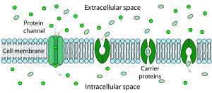
Facilitated diffusion in the cell membrane, showing ion channels (left) and carrier proteins (three on the right).
Facilitated diffusion is the passage of molecules or ions across a biological membrane through specific transport proteins and requires no energy input. Facilitated diffusion is used especially in the case of large polar molecules and charged ions; once such ions are dissolved in water they cannot diffuse freely across cell membranes due to the hydrophobic nature of the fatty acid tails of the phospholipids that make up the bilayers.
The type of carrier proteins used in facilitated diffusion is slightly different from those used in active transport. They are still transmembrane carrier proteins, but these are gated transmembrane channels, meaning they do not internally translocate, nor require ATP to function. The substrate is taken in one side of the gated carrier, and without using ATP the substrate is released into the cell. Facilitated diffusion does not require the use of ATP as facilitated diffusion, like simple diffusion, transports molecules or ions along their concentration gradient.[15]
Osmosis is the passive diffusion of water across a cell membrane from an area of high concentration to an area of low concentration. Since Osmosis is a passive process, like facilitated diffusion and simple diffusion, it does not require the use of ATP. Osmosis is important in regulating the balance of water and salt within cells, thus it plays a critical role in maintaining homeostasis.[16]Aquaporins are integral membrane proteins that allow for the rapid passage of water and glycerol through membranes. The aquaporin monomers consist of six transmembrane alpha-helix domains and these monomers can assemble to form the aquaporin proteins. As four of these monomers come together to form the aquaporin protein, it is known as a homotetramer, meaning it is made up of four identical subunits.[17][18] All aquaporins are tetrameric membrane integral proteins, and the water passes through each individual monomer channel rather than between all of the four channels. Since aquaporins are transmembrane channels for the diffusion of water, the channels that make up the aquaporin are typically lined with hydrophilic side chains to allow water to pass through.
Reverse transport, or transporter reversal, is a phenomenon in which the substrates of a membrane transport protein are moved in the opposite direction to that of their typical movement by the transporter.[19][20][21][22][23] Transporter reversal typically occurs when a membrane transport protein is phosphorylated by a particular protein kinase, which is an enzyme that adds a phosphate group to proteins.[19][20]
Nonribosomally synthesized channels such as gramicidin
Holins; which function in export of enzymes that digest bacterial cell walls in an early step of cell lysis.
Facilitated diffusion occurs in and out of the cell membrane via channels/pores and carriers/porters.
Note:
Channels:
Channels are either in open state or closed state. When a channel is opened with a slight conformational switch, it is open to both environment simultaneously (extracellular and intracellular)
This picture represents symport. The yellow triangle shows the concentration gradient for the yellow circles while the green triangle shows the concentration gradient for the green circles and the purple rods are the transport protein bundle. The green circles are moving against their concentration gradient through a transport protein which requires energy while the yellow circles move down their concentration gradient which releases energy. The yellow circles produce more energy through chemiosmosis than what is required to move the green circles so the movement is coupled and some energy is cancelled out. One example is the lactose permease which allows protons to go down its concentration gradient into the cell while also pumping lactose into the cell.Pores:
Pores are continuously open to these both environment, because they do not undergo conformational changes. They are always open and active.
2: Electrochemical potential-driven transporters
Also named carrier proteins or secondary carriers.
The picture represents uniport. The yellow triangle shows the concentration gradient for the yellow circles and the purple rods are the transport protein bundle. Since they move down their concentration gradient through a transport protein, they can release energy as a result of chemiosmosis. One example is GLUT1 which moves glucose down its concentration gradient into the cell.Excitatory amino acid transporters (EAATs)
This picture represents antiport. The yellow triangle shows the concentration gradient for the yellow circles while the blue triangle shows the concentration gradient for the blue circles and the purple rods are the transport protein bundle. The blue circles are moving against their concentration gradient through a transport protein which requires energy while the yellow circles move down their concentration gradient which releases energy. The yellow circles produce more energy through chemiosmosis than what is required to move the blue circles so the movement is coupled and some energy is cancelled out. One example is the sodium-proton exchanger which allows protons to go down their concentration gradient into the cell while pumping sodium out of the cell.F-type ATPase; ("F" related to factor), including: mitochondrial ATP synthase, chloroplast ATP synthase1
3.B: Decarboxylation-driven transporters
3.C: Methyltransfer-driven transporters
3.D: Oxidoreduction-driven transporters
3.E: Light absorption-driven transporters, such as rhodopsin
4: Group translocators
The group translocators provide a special mechanism for the phosphorylation of sugars as they are transported into bacteria (PEP group translocation)
5: Electron carriers
The transmembrane electron transfer carriers in the membrane include two-electron carriers, such as the disulfide bond oxidoreductases (DsbB and DsbD in E. coli) as well as one-electron carriers such as NADPH oxidase. Often these redox proteins are not considered transport proteins.
Relevant Examples
GLUT 1
Every carrier protein, especially within the same cell membrane, is specific to one type or family of molecules. GLUT1 is a named carrier protein found in almost all animal cell membranes that transports glucose across the bilayer. This protein is a uniporter, meaning it transports glucose along its concentration in a singular direction. It is an integral membrane protein carrier with a hydrophilic interior, which allows it to bind to glucose. As GLUT 1 is a type of carrier protein, it will undergo a conformational change to allow glucose to enter the other side of the plasma membrane.[24] GLUT 1 is commonly found in the red blood cell membranes of mammals.[25]
Sodium/Potassium Channels
While there are many examples of channels within the human body, two notable ones are sodium and potassium channels. Potassium channels are typically involved in the transport of potassium ions across the cell membrane to the outside of the cell, which helps maintain the negative membrane potential of cells. As there are more potassium channels than sodium channels, more potassium flows out of the cell than sodium into a cell, thus why the membrane potential is negative. Sodium channels are typically involved in the transport of sodium ions across the cell membrane into the cell. These channels are commonly associated with excitable neurons, as an influx of sodium can trigger depolarization, which in turn propagates an action potential.[26] As these proteins are types of channel proteins, they do not undergo a change of conformation after binding their respective substrates.
Other Examples
Other specific carrier proteins also help the body function in important ways. Cytochromes operate in the electron transport chain as carrier proteins for electrons.[12]
Pathology
A number of inherited diseases involve defects in carrier proteins in a particular substance or group of cells. Cysteinuria (cysteine in the urine and the bladder) is such a disease involving defective cysteine carrier proteins in the kidney cell membranes. This transport system normally removes cysteine from the fluid destined to become urine and returns this essential amino acid to the blood. When this carrier malfunctions, large quantities of cysteine remain in the urine, where it is relatively insoluble and tends to precipitate. This is one cause of urinary stones.[27] Some vitamin carrier proteins have been shown to be overexpressed in patients with malignant disease. For example, levels of riboflavin carrier protein (RCP) have been shown to be significantly elevated in people with breast cancer.[28]
^Hediger, Matthias A.; Romero, Michael F.; Peng, Ji-Bin; Rolfs, Andreas; Takanaga, Hitomi; Bruford, Elspeth A. (February 2004). "The ABCs of solute carriers: physiological, pathological and therapeutic implications of human membrane transport proteinsIntroduction". Pflügers Archiv: European Journal of Physiology. 447 (5): 465–468. doi:10.1007/s00424-003-1192-y. ISSN0031-6768. PMID14624363. S2CID1866661.
^ abPerland, Emelie; Fredriksson, Robert (March 2017). "Classification Systems of Secondary Active Transporters". Trends in Pharmacological Sciences. 38 (3): 305–315. doi:10.1016/j.tips.2016.11.008. ISSN1873-3735. PMID27939446.
^Huang, Y; Anderle, P; Bussey, KJ; Barbacioru, C; Shankavaram, U; Dai, Z; Reinhold, WC; Papp, A; Weinstein, JN; Sadée, W (15 June 2004). "Membrane transporters and channels: role of the transportome in cancer chemosensitivity and chemoresistance". Cancer Research. 64 (12): 4294–301. doi:10.1158/0008-5472.CAN-03-3884. PMID15205344. S2CID2765236.
^Sadava, David, et al. Life, the Science of Biology, 9th Edition. Macmillan Publishers, 2009. ISBN1-4292-1962-9. p. 119.
^Cooper, Geoffrey (2009). The Cell: A Molecular Approach. Washington, DC: ASM Press. p. 62. ISBN9780878933006.
^Thompson, Liz A. Passing the North Carolina End of Course Test for Biology. American Book Company, Inc. 2007. ISBN1-59807-139-4. p. 97.
^Assmann, Sarah (2015). "Solute Transport". In Taiz, Lincoln; Zeiger, Edward (eds.). Plant Physiology and Development. Sinauer. p. 151.
^Sadava, David, Et al. Life, the Science of Biology, 9th Edition. Macmillan Publishers, 2009. ISBN1-4292-1962-9. p. 119.
^Kasatkina LA, Borisova TA (November 2013). "Glutamate release from platelets: exocytosis versus glutamate transporter reversal". The International Journal of Biochemistry & Cell Biology. 45 (11): 2585–2595. doi:10.1016/j.biocel.2013.08.004. PMID23994539.
^Cooper, Geoffrey M. (2000), "Transport of Small Molecules", The Cell: A Molecular Approach. 2nd edition, Sinauer Associates, retrieved 2023-11-22
^Sherwood, Lauralee. 7th Edition. Human Physiology. From Cells to Systems. Cengage Learning, 2008. p. 67
^Rao, PN, Levine, E et al. Elevation of Serum Riboflavin Carrier Protein in Breast Cancer. Cancer Epidemiol Biomarkers Prev. Volume 8 No 11. pp. 985–990
Anderle, P., Barbacioru,C., Bussey, K., Dai, Z., Huang, Y., Papp, A., Reinhold, W., Sadee, W., Shankavaram, U., & Weinstein, J. (2004). Membrane Transporters and Channels: Role of the Transportome in Cancer Chemosensitivity and Chemoresistance. Cancer Research, 54, 4294-4301.