
| Pterygoid processes of the sphenoid | |
|---|---|
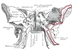
 Sphenoid bone, upper surface. | |
 Sphenoid bone, anterior and inferior surfaces. | |
| Details | |
| Identifiers | |
| Latin | processus pterygoideus ossis sphenoidalis |
| TA98 | A02.1.05.042 |
| TA2 | 628 |
| FMA | 54682 |
| Anatomical terms of bone | |
The pterygoid processes of the sphenoid (from Greek pteryx, pterygos, "wing"), one on either side, descend perpendicularly from the regions where the body and the greater wings of the sphenoid bone unite.
Each process consists of a medial pterygoid plate and a lateral pterygoid plate, the latter of which serve as the origins of the medial and lateral pterygoid muscles. The medial pterygoid, along with the masseter allows the jaw to move in a vertical direction as it contracts and relaxes. The lateral pterygoid allows the jaw to move in a horizontal direction during mastication (chewing). Fracture of either plate are used in clinical medicine to distinguish the Le Fort fracture classification for high impact injuries to the sphenoid and maxillary bones.
The superior portion of the pterygoid processes are fused anteriorly; a vertical groove, the pterygopalatine fossa, descends on the front of the line of fusion. The plates are separated below by an angular cleft, the pterygoid notch, the margins of which are rough for articulation with the pyramidal process of the palatine bone.
The two plates diverge behind and enclose between them a V-shaped fossa, the pterygoid fossa, which contains the medial pterygoid muscle and the tensor veli palatini.
Above this fossa is a small, oval, shallow depression, the scaphoid fossa, which gives origin to the tensor veli palatini.
The anterior surface of the pterygoid process is broad and triangular near its root, where it forms the posterior wall of the pterygopalatine fossa and presents the anterior orifice of the pterygoid canal.
In many mammals it remains as a separate bone called the pterygoid bone.
Its name is Greek for "resembling a fin or wing", from its shape.
YouTube Encyclopedic
-
1/5Views:177 0408 807153 9263 32010 723
-
Sphenoid Bone - Definition, Location & Function - Human Anatomy | Kenhub
-
Infratemporal Fossa LO - Pterygoid Plates
-
SPHENOID BONE
-
Sphenoid 2019
-
Sphenoid
Transcription
Hello, again, everyone. It’s Matt from Kenhub. And in this tutorial, we will discuss the sphenoid bone. The sphenoid bone is the most complex bone of the human body. Because of its shape, it is also known as the wasp bone. It makes up most of the middle part of the base of the skull and contributes to the floor of the middle cranial fossa. There are four main parts to the sphenoid bone – the body, the lesser and greater wings, and the pterygoid processes. The body is the most centrally positioned portion. Anteriorly, it contributes to the nasal cavity. Laterally, it builds the medial wall of the optic canal. Superiorly, the body forms the sella tursica, the hypophyseal fossa, and the dorsum sellae. They contain the anterior and posterior glenoid processes, respectively. The clivus slopes posterior to the body. The sphenoidal sinuses are located in the sphenoid body behind the nasal cavity and are divided by a septum. The lesser wings arise supralaterally from the sphenoid body where they form the optic canal which contains the optic nerve and the ophthalmic artery. The inferior surface participates in the lateral margin of the orbit. While the superior surface forms part of the cranial cavity. The greater wings arise posterolaterally from the body. Their lateral surfaces form the infratemporal surfaces. Their anterior surfaces make up part of the posterior aspects of the lateral wall of the orbit. They contain two important openings near their roots, the foramen rotundum which houses the maxillary nerve and the foramen ovale containing the mandibular nerve and the accessory meningeal artery. The foramen spinosum which is the opening for the middle meningeal vessel and the spinous nerve lies at the posterior margin of the greater wings. Between the body, lesser and greater wings is a large opening known as the superior orbital fissure where numerous nerves and vessels pass through including the superior ophthalmic vein, ophthalmic nerve and its branches, abducens nerve, oculomotor nerve, and the trochlear nerve. The pterygoid processes are extensions of the basal surface of the sphenoid body. The processes contain two canals known as the pterygoid canal housing the major petrosal nerve, deep petrosal nerve and vessels of pterygoid canal, and the palatovaginal or pharangeal canal which holds the pharangeal nerve. A hamulus extends bilaterally from each medial pterygoid plate. The sphenoid bone has a common border with the frontal bone via the sphenofrontal suture, the parietal bone via the sphenoparietal suture, the squamous part of the temporal bone via the sphenosquamosal suture, and the occipital bone via the spheno-occipital suture. As the sphenoid and occipital bone fuse during puberty, the sphenooccipital suture disappears by the age of 25. The body and lesser wings of the sphenoid bone matures through classic endochondral ossification, whereas the pterygoid processes undergo intramembranous ossification. The development of the greater wings of the sphenoid bone are exceptional, since they are the only bony structures of the skull which go through both endochondral and intramembranous ossification. This video is more fun than reading a textbook, right? If you want more videos, interactive quizzes, articles, and an atlas of human anatomy, click on the “Take me to Kenhub” button. It is time to say goodbye to your old textbooks and say hello to your new anatomy learning partner, Kenhub! See you there! https://www.kenhub.com
Medial pterygoid plate
The medial pterygoid plate (or medial pterygoid lamina) of the sphenoid bone is a horse-shoe shaped process that arises from its underside.
It is narrower and longer than the lateral pterygoid plate and curves lateralward at its lower extremity into a hook-like process, the pterygoid hamulus, around which the tendon of the tensor veli palatini glides.
The lateral surface of this plate forms part of the pterygoid fossa, the medial surface constitutes the lateral boundary of the choana or posterior aperture of the corresponding nasal cavity.
Superiorly the medial plate is prolonged on to the under surface of the body as a thin lamina, named the vaginal process, which articulates in front with the sphenoidal process of the palatine and behind this with the ala (wing) of the vomer.
The angular prominence between the posterior margin of the vaginal process and the medial border of the scaphoid fossa is named the pterygoid tubercle, and immediately above this is the posterior opening of the pterygoid canal.
On the under surface of the vaginal process is a furrow, which is converted into a canal by the sphenoidal process of the palatine bone, for the transmission of the pharyngeal branch of the internal maxillary artery and the pharyngeal nerve from the sphenopalatine ganglion.
The pharyngeal aponeurosis is attached to the entire length of the posterior edge of the medial plate, and the constrictor pharyngis superior takes origin from its lower third.
Projecting backward from near the middle of the posterior edge of this plate is an angular process, the processus tubarius, which supports the pharyngeal end of the Eustachian tube.
The anterior margin of the plate articulates with the posterior border of the vertical part of the palatine bone.
In many animals it is a separate bone called the pterygoid bone.
Lateral pterygoid plate
| Lateral pterygoid plate | |
|---|---|
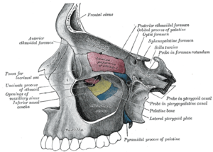 Medial wall of left orbit. (Lateral pterygoid plate labeled at bottom right.) | |
| Details | |
| Identifiers | |
| Latin | lamina lateralis processus pterygoidei |
| TA98 | A02.1.05.042 |
| TA2 | 628 |
| FMA | 54682 |
| Anatomical terms of bone | |
The lateral pterygoid plate of the sphenoid (or lateral lamina of pterygoid process) is broad, thin, and everted and forms the lateral part of a horseshoe like process that extends from the inferior aspect of the sphenoid bone, and serves as the origin of the lateral pterygoid muscle, which functions in allowing the mandible to move in a lateral and medial direction, or from side-to-side.
Its lateral surface forms part of the medial wall of the infratemporal fossa, and gives attachment to the lateral pterygoid muscle; its medial surface forms part of the pterygoid fossa, and gives attachment to the medial pterygoid muscle. Posterior edge is sharp, and often has sharp projection - pterygospinous process (Civinini process).
References
![]() This article incorporates text in the public domain from page 151 of the 20th edition of Gray's Anatomy (1918)
This article incorporates text in the public domain from page 151 of the 20th edition of Gray's Anatomy (1918)
External links
- "Anatomy diagram: 34257.000-1". Roche Lexicon - illustrated navigator. Elsevier. Archived from the original on 2012-07-22.
- Anatomy figure: 22:4b-05 at Human Anatomy Online, SUNY Downstate Medical Center
- "Anatomy diagram: 25420.000-1". Roche Lexicon - illustrated navigator. Elsevier. Archived from the original on 2015-02-26.