
| Facial canal | |
|---|---|
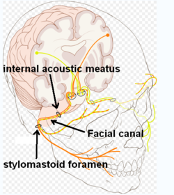 Route of facial nerve, with facial canal labeled | |
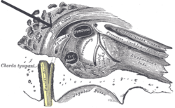 View of the inner wall of the tympanum. (Facial canal visible in upper left; promontory labeled at center) | |
| Details | |
| System | skeletal |
| Nerve | facial nerve (CN VII) |
| Identifiers | |
| Latin | canalis nervi facialis, canalis facialis |
| TA98 | A02.1.06.009 |
| TA2 | 688 |
| FMA | 54952 |
| Anatomical terminology | |
The facial canal (also known as the Fallopian canal) is a Z-shaped canal in the temporal bone of the skull. It extends between the internal acoustic meatus and stylomastoid foramen. It transmits the facial nerve (CN VII) (after which it is named).
YouTube Encyclopedic
-
1/5Views:259 253396 738137 86580 079349 677
-
The Facial Nerve (CNVII): Animated Review
-
Facial Nerve Anatomy Simplified
-
Facial nerve: branches and course (preview) - Human Neuroanatomy | Kenhub
-
Facial nerve - Origin, Function, Pathway & Branches | Anatomy Tutorial
-
Neurology | Facial Nerve: Cranial Nerve VII
Transcription
Anatomy
The facial canal gives passage to the facial nerve (CN VII) (hence the name).[1][verification needed][better source needed] Its proximal opening is at the internal auditory meatus; its distal opening is the stylomastoid foramen. In humans, the canal is approximately 3 cm long, making it the longest bony canal of a nerve in the human body.[2][verification needed][better source needed] It is located within the middle ear region.[verification needed][better source needed]
The facial nerve gives rise to three nerves while passing through the canal: the greater petrosal nerve, nerve to stapedius, and the chorda tympani.[3]
Structure
Horizontal part
The proximal portion of the facial canal is termed the horizontal part. It commences at the introitus of facial canal at the distal end of the internal auditory meatus. The horizontal part is further subdivided into two crura: the proximal/medial[4] anteriolaterally[5] directed medial crus (or labyrinthine segment[5]), and the distal/lateral[4] posteriolaterally[5] directed lateral crus (or tympanic segment[5]); the two crura meet at a sharp angle at the genu of facial canal (geniculum canalis facialis[6]) where the geniculate ganglion is situated (at the genu, the greater petrosal nerve leaves the facial canal through the hiatus of the facial canal).[4]
Descending part
The lateral crus of horizontal part ends by turning sharply inferior-ward, commencing the distal-most descending part (or mastoid segment[5]) of facial canal which passes vertically inferior-ward, ending distally at the stylomastoid foramen. The descending part presents two openings through each of which a branch of the facial nerve passes: the nerve to stapedius enters the canaliculus for nerve to stapedius, and the chorda tympani enters the posterior canaliculus of chorda tympani (canaliculus chordae tympani, or iter chordae posterius[7]).[8]
Relations
The labyrinthine segment is situated superior to cochlea.[5]
The canal traverses the medial wall of the tympanic cavity superior to the oval window;[citation needed] here, the prominence of the facial canal (or prominence of the aqueduct of Fallopius) upon the medial wall indicates the position of the superior portion of the facial canal.[9]: 745 The canal then curves nearly vertically inferior-ward along the posterior wall.[citation needed] The tympanic segment is closely related to the posterior and medial walls of the tympanic cavity; it passes superior to the oval window and inferior to the lateral semicircular canal.[5]
Clinical significance
The facial canal may be interrupted in some people. This may lead to the facial nerve being split into 2 or 3 fibres, or it may be poorly formed or congenitally absent on one side.[2]
History
The facial canal was first described by Gabriele Falloppio.[10] This is why it may also be known as the Fallopian canal.[10]
Gallery
-
 Lateral head anatomy detail. Facial nerve dissection.
 -
 Tympanic cavity. Facial canal. Internal carotid artery.
 -
 Coronal section of right temporal bone. Prominence of the facial canal labeled at top, fourth from the left.

See also
References
- ^ Nager, George T.; Proctor, Bruce (1991-06-01). "Anatomie Variations and Anomalies Involving the Facial Canal". Otolaryngologic Clinics of North America. 24 (3): 531–553. doi:10.1016/S0030-6665(20)31114-2. ISSN 0030-6665. PMID 1762775.
- ^ a b Weiglein AH (June 1996). "Postnatal development of the facial canal. An investigation based on cadaver dissections and computed tomography". Surgical and Radiologic Anatomy. 18 (2): 115–23. doi:10.1007/BF01795229. PMID 8782317. S2CID 25764734.
- ^ Moore, Keith L.; Dalley, Arthur F.; Agur, Anne M. R. (2018). Clinically Oriented Anatomy (8th ed.). Wolters Kluwer. p. 1077. ISBN 978-1-4963-4721-3.
- ^ a b c "horizontal part of facial canal". TheFreeDictionary.com. Retrieved 2023-07-29.
- ^ a b c d e f g Shin KJ, Gil YC, Lee JY, Kim JN, Song WC, Koh KS (October 2014). "Three-dimensional study of the facial canal using microcomputed tomography for improved anatomical comprehension". Anatomical Record. 297 (10): 1808–16. doi:10.1002/ar.22977. PMID 24990524. S2CID 205411993.
- ^ "genu of facial canal". TheFreeDictionary.com. Retrieved 2023-07-29.
- ^ "posterior canaliculus of chorda tympani". TheFreeDictionary.com. Retrieved 2023-07-29.
- ^ "descending part of facial canal". TheFreeDictionary.com. Retrieved 2023-07-29.
- ^ Standring, Susan (2020). Gray's Anatomy: The Anatomical Basis of Clinical Practice (42th ed.). New York. ISBN 978-0-7020-7707-4. OCLC 1201341621.
{{cite book}}: CS1 maint: location missing publisher (link) - ^ a b Abing W, Rauchfuss A (2005). "Fetal development of the tympanic part of the facial canal". European Archives of Oto-Rhino-Laryngology. 243 (6): 374–377. doi:10.1007/bf00464645. PMID 3566620. S2CID 12712839.