
| Dorsum sellae | |
|---|---|
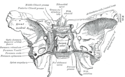 Sphenoid bone. Upper surface. (Dorsum sellae is labeled in the white portion in the center.) | |
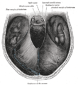
 Base of the skull. Upper surface. (Dorsum sellae is not labeled, but sphenoid bone is in yellow, and Dorsum sellae would horizontally be at the exact center, and vertically at the level of the posterior clinoid process.) | |
| Details | |
| Identifiers | |
| Latin | dorsum sellae, dorsum sella turcica |
| TA98 | A02.1.05.010 |
| TA2 | 594 |
| FMA | 54718 |
| Anatomical terms of bone | |
The dorsum sellae is part of the sphenoid bone in the skull. Together with the basilar part of the occipital bone it forms the clivus.
In the sphenoid bone, the anterior boundary of the sella turcica is completed by two small eminences, one on either side, called the middle clinoid processes, while the posterior boundary is formed by a square-shaped plate of bone, the dorsum sellae, ending at its superior angles in two tubercles, the posterior clinoid processes, the size and form of which vary considerably in different individuals.
YouTube Encyclopedic
-
1/3Views:55 896726628
-
Sphenoid Bone - Anatomy, Function and Definition - Human Anatomy | Kenhub
-
DORSUM SELLA MENINGIOMA-microsurgical removal-dr suresh dugani/ HUBLI/ KARNATAK /INDIA
-
Endocrine Lab 2, Part 1.wmv
Transcription
Hello, again, everyone. It’s Matt from Kenhub. And in this tutorial, we will discuss the sphenoid bone. The sphenoid bone is the most complex bone of the human body. Because of its shape, it is also known as the wasp bone. It makes up most of the middle part of the base of the skull and contributes to the floor of the middle cranial fossa. There are four main parts to the sphenoid bone – the body, the lesser and greater wings, and the pterygoid processes. The body is the most centrally positioned portion. Anteriorly, it contributes to the nasal cavity. Laterally, it builds the medial wall of the optic canal. Superiorly, the body forms the sella tursica, the hypophyseal fossa, and the dorsum sellae. They contain the anterior and posterior glenoid processes, respectively. The clivus slopes posterior to the body. The sphenoidal sinuses are located in the sphenoid body behind the nasal cavity and are divided by a septum. The lesser wings arise supralaterally from the sphenoid body where they form the optic canal which contains the optic nerve and the ophthalmic artery. The inferior surface participates in the lateral margin of the orbit. While the superior surface forms part of the cranial cavity. The greater wings arise posterolaterally from the body. Their lateral surfaces form the infratemporal surfaces. Their anterior surfaces make up part of the posterior aspects of the lateral wall of the orbit. They contain two important openings near their roots, the foramen rotundum which houses the maxillary nerve and the foramen ovale containing the mandibular nerve and the accessory meningeal artery. The foramen spinosum which is the opening for the middle meningeal vessel and the spinous nerve lies at the posterior margin of the greater wings. Between the body, lesser and greater wings is a large opening known as the superior orbital fissure where numerous nerves and vessels pass through including the superior ophthalmic vein, ophthalmic nerve and its branches, abducens nerve, oculomotor nerve, and the trochlear nerve. The pterygoid processes are extensions of the basal surface of the sphenoid body. The processes contain two canals known as the pterygoid canal housing the major petrosal nerve, deep petrosal nerve and vessels of pterygoid canal, and the palatovaginal or pharangeal canal which holds the pharangeal nerve. A hamulus extends bilaterally from each medial pterygoid plate. The sphenoid bone has a common border with the frontal bone via the sphenofrontal suture, the parietal bone via the sphenoparietal suture, the squamous part of the temporal bone via the sphenosquamosal suture, and the occipital bone via the spheno-occipital suture. As the sphenoid and occipital bone fuse during puberty, the sphenooccipital suture disappears by the age of 25. The body and lesser wings of the sphenoid bone matures through classic endochondral ossification, whereas the pterygoid processes undergo intramembranous ossification. The development of the greater wings of the sphenoid bone are exceptional, since they are the only bony structures of the skull which go through both endochondral and intramembranous ossification. This video is more fun than reading a textbook, right? If you want more videos, interactive quizzes, articles, and an atlas of human anatomy, click on the “Take me to Kenhub” button. It is time to say goodbye to your old textbooks and say hello to your new anatomy learning partner, Kenhub! See you there! https://www.kenhub.com
Additional images
-
 Tentorium cerebelli from above.
 -
 Dorsum sellae

References
![]() This article incorporates text in the public domain from page 147 of the 20th edition of Gray's Anatomy (1918)
This article incorporates text in the public domain from page 147 of the 20th edition of Gray's Anatomy (1918)
External links
- "Anatomy diagram: 34257.000-2". Roche Lexicon - illustrated navigator. Elsevier. Archived from the original on 2013-06-22.
- Anatomy photo:22:os-0912 at the SUNY Downstate Medical Center
This human musculoskeletal system article is a stub. You can help Wikipedia by expanding it. |