
 | |
| Identifiers | |
|---|---|
| |
| CAS Number |
|
| PubChem CID | |
| IUPHAR/BPS | |
| ChemSpider | |
| UNII |
|
| ChEMBL | |
| CompTox Dashboard (EPA) | |
| Chemical and physical data | |
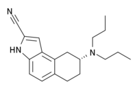
| Formula | C19H25N3 |
| Molar mass | 295.430 g·mol−1 |
| 3D model (JSmol) | |
| |
| |
| (verify) | |
U-92,016-A is a psychoactive drug and research chemical used in scientific studies. It acts as a potent, high efficacy, and selective 5-HT1A receptor full agonist with a long duration of action.[1][2] It has been suggested that it could be developed as an anxiolytic or antidepressant drug.[1]
YouTube Encyclopedic
-
1/1Views:1 730 857
-
Parts of a cell
Transcription
Let's talk a little bit about the structure of the cell. I've done a lot of videos where we deal with things that go on inside of them, but I haven't done one where we just talk about the entire structure of them. So a good place to start is-- let me just draw the membrane. And the cellular membrane is a good place to start because this is what separates the cell from the outside world, and to a large degree, it kind of defines the cell. It defines it as this very, very small compartment. That's where the word, "cell" came from. So let me label that. Cellular membrane. And all cells have a cellular membrane. Now if we think about maybe the most important thing that defines a cell, you've probably seen in the DNA videos and we're going to talk about translation and transcription and all of that, that what defines what a living organism is, is its DNA. So all cells have DNA inside of it. And I won't go into the details of how DNA defines what an organism is. I've done that in some detail on the DNA videos. But all cells have DNA. This is more of an anatomy of a cell video than necessarily the function, but we'll go into the function because we need to know what these different parts do. So this is, right here, this is the DNA. And it's in its chromatin form here. There's also little proteins here. Not in all organisms, but we're going to stick to eukaryotes, and I'll talk a little bit about the difference between eukaryotes and prokaryotes in a second. But we have DNA. As I've drawn this cell right now, this is pretty much any cell, and any animal or plant or whatever kingdom could look like this. I haven't drawn a lot of the details. I've just drawn the DNA and the cellular membrane. Now here's kind of the first major division in the living world, or at least from our point of view, or it seemed obvious, is that some cells have a membrane around the DNA. So they'll have a membrane around the DNA that separates the DNA and the chromatin and everything that makes up the stuff within the DNA, separates that from the rest of the cell, and this is called a nucleus. This is called a nucleus. And I said that's a major division because when some people looked at some cells and they saw a nucleus and other cells and they didn't see a nucleus, they said, hey, this is a good way to classify organisms. So they called the things that had nucleuses, eukaryotes. These have a nucleus. So as I've drawn this cell right here, it is a eukaryote. Now, if you do not have a nucleus, you are dealing with a prokaryote. No nucleus. And examples of prokaryotes, the two big groups of them, are bacteria and Archaea. Now, Archaea are really interesting. We know very little about them. They were originally thought to be types of bacteria, but now people are realizing that they're this whole completely other group, and we've actually observed a very small subset of them, so it's a very fascinating group. And it actually turns out that, evolutionarily speaking, you shouldn't make this division first. It actually makes more sense to divide things into eukaryotes, I'll just write that Euk, bacteria and Archaea. You don't want to do this division first. There are actually three separate groups that you want to start off with. We'll talk more about this in future videos. But if you want to say who has a nucleus? Well, eukaryotes have a nucleus by definition. Who does not have a nucleus? Well, the bacteria and the Archaea do not have nuclei, just like that. But I'm going to focus on eukaryotes because they tend to be a little bit more complex. They tend to be larger. And most of what we talk about, at least in the videos so far, are dealing with eukaryotes. Eukaryotes include plants, animals-- we're animals, at least I am-- animals and fungi, and there are other groups within eukaryotes, but these are the ones that we normally deal with in our everyday world. But let's go back to looking at the anatomy of the cell. So we have our DNA. We know that it gets transcribed into mRNA, That mRNA leaves the nucleus, and it gets translated into proteins at the ribosomes. So the ribosomes are these little complexes that could be floating all over the cell, and we'll see in a second that they can also be attached to these other membrane structures. So this is a ribosome. And if all this talk of DNA transcription into mRNA and mRNA leaving the nucleus and traveling to the ribosome to be translated into proteins makes no sense to you, there are several videos where I go into that in detail. But what I want to do is just focus on all of the different parts to kind of give a big picture of things. So ribosomes are where mRNA that gets transcribed inside the nucleus from DNA, where it gets translated into proteins. So you can kind of view them as the place where information actually turns into proteins, which then can be used anywhere else in the cell. And these ribosomes, they're made up of proteins, and actually they're made up RNA. And so one question is, well, where are the pieces of the ribosomes made? Well, some are made by proteins, which might be made in other ribosomes. But some of it, the mRNA, ribosomes, you can kind of view them as this big mess, if you were to look at it in detail. There's some protein there. And I'm not drawing it that realistically, but then you have some mRNA tied in with the protein, and the mRNA isn't used for actual information purposes like it normally is when it goes from DNA to the ribosome. Within the ribosome itself, ribosomal RNA is actually used as part of the structure. It actually helps the ribosome function as a ribosome. So it's actually part of the ribosome. And all of that gets built in a part of the nucleus called the nucleolus or the nucleole. Let me write that down. So this right here, this is interesting right here. This is the nucleolus or the nucleole. And it isn't a separate organelle, and it's not separated by a membrane, but it shows up in a microscope. So when people first saw it, they said, hey, there's like a bundle there. That must be like the core of the nucleus or something. But it turns out what it is, it's the densely packed-- you've got DNA and RNA, and that's actually where the ribosomal RNA, the stuff that makes up the ribosomes, is actually produced. But it's so dense that it shows up on a microscope, and that's why people decided to name it something different. But it is not membrane bound. It's not an organelle within an organelle. It's just densely packed proteins and ribosomal RNA, and it's where ribosomal RNA gets produced. So anyway, we're at the ribosome. That's where proteins get produced. But if the ribosomes are just floating around, if they're free ribosomes, then those proteins will just-- once they're produced at the ribosome, they'll just float around here in the fluid inside the cell that we call cytosol. But what if we wanted to produce proteins that are supposed to end up, maybe in the membrane of the cell, or maybe outside of the cell itself? Cells produce things that are used by other cells, that are used by the rest of the body. And here we have to go to proteins that are attached to this membrane. You can kind of view it as a bunch of tunnels. Let me see how well I can draw that. I'm going to draw it very roughly. You have this thing called the endoplasmic reticulum. Endoplasmic reticulum. You can just view it as a bunch of tunnels like that. Endoplasmic reticulum. And they eventually lead to something called Golgi bodies, named after Mr. Golgi himself. So I'll do the endoplasmic reticulum in yellow and I'll do the Golgi bodies in green maybe, just like that. I'll tell you in a second what they are. So what's going on? This is kind of a big kind of stack, or you can kind of view it as a bunch of folded membranes together. And some ribosomes are actually attached to this part, which I call the endoplasmic reticulum. So we have ribosomes attached. Some of them are free, some of them are attached. And let me write my labels down. So this right here, and we use the space over here, this big flap of convoluted membrane, that's the endoplasmic reticulum. It's fun to say. Endoplasmic reticulum. Maybe a good name for a band. Endoplasmic reticulum, and the parts that have ribosomes attached to it are called the rough endoplasmic reticulum. Maybe even a better name for a band. So right here, where I have attached ribosomes, so these ribosomes are attached right here, this is the rough endoplasmic reticulum, or maybe the rough ER. The rough ER, ER standing for endoplasmic reticulum. And then where we don't have any ribosomes attached, that's the smooth endoplasmic reticulum. So right here, that is this smooth endoplasmic reticulum. And I'm going to tell you in a second what this is, but we can keep following the membranes. Eventually we get the Golgi bodies. And what happens is, I gave you a hint. In our free ribosomes, mRNA gets there, gets translated into proteins, and the proteins then just float around in the cytosol. But what if we want proteins that should end up in membranes or that end up outside of the cell? And that's where the endoplasmic reticulum and the Golgi bodies come into play. Because now what we can do is, we have mRNA coming outside of the nucleus, and it can attach to the ribosomes or it can be translated by the ribosomes in the rough ER. And what happens here is your mRNA comes here and it's getting-- and I drew that arrow very small-- it's getting translated on the outside of the endoplasmic reticulum, but as the protein gets produced, it's getting pushed into the inside of the endoplasmic reticulum. And when I say the inside of the endoplasmic reticulum, I'm talking about this area. I'm coloring it in. This is the inside of the endoplasmic reticulum. And so the proteins will get pushed into the endoplasmic reticulum. The ones that are to be used outside of the cytosol, outside of the cell or maybe in the cell's membrane. So proteins will end up here. That's why those ribosomes are on the membrane, because they can translate things that are outside of the endoplasmic reticulum, but as the proteins get produced, the amino acid chain ends up inside of it. Let me make a blow-up of that because I think that'll be useful. So let me draw-- let's say this is the membrane of the endoplasmic reticulum and then you have ribosomes attached to it. Let's say that's a ribosome on the endoplasmic reticulum. And is this is going to be the rough endoplasmic reticulum. And what you can have is mRNA coming into one side of it. mRNA can kind of come in through here. Maybe it's going in that direction. It's getting translated into the proteins. But then the protein, as the amino acid chain is built up, will pop out on this end of the membrane. Remember, this is our membrane of our endoplasmic reticulum. So even though the mRNA is on the outside, because the ribosome is attached to it, the protein can show up on the inside. So once the protein is built, maybe then it folds all up, you know, a protein is just a folded chain of amino acids, it can travel through the endoplasmic reticulum. And it travels through it. It travels through the smooth endoplasmic reticulum all the way until it gets to the Golgi bodies. And all sorts of other things happen. I'm oversimplifying things, but I just want to give you a sense of what everything in the cell does. And once the protein travels to the Golgi bodies and they get ready for traveling outside of the cell, or maybe traveling to the cell's membrane, they'll actually bud out of the Golgi bodies. So let's say this same protein, when it gets to the Golgi bodies-- remember it's inside of the Golgi body, so let me draw the membrane of the Golgi body right there. The protein might end up there. It's just a big chain of amino acids. And then it will bud out. So let's say it looks like that, and then maybe the next step it will look something like. It will look like that. And the very next step, you could imagine, will look something like this, where it's actually completely budded out. It's popped out a little of the membrane of the Golgi body with it. So now the protein is surrounded by its own little membrane. So let's think about what happened. We had DNA transcribed into mRNA. mRNA goes to a ribosome that's attached to the endoplasmic reticulum. It is translated into a protein that travels through the endoplasmic reticulum. First, the rough, where all the ribosomes are, then this smooth. The smooth has other functions. It also helps produce hormones and other fatty compounds, but I won't go into detail there. But it just travels. It connects to the Golgi bodies. Then the Golgi bodies, the proteins can bud off and take a little bit of the membrane with it. And this idea of something being surrounded by a membrane and just traveling in the cell, so maybe the protein now looks like this. I'm enlarging it. Maybe the protein is there and then it took a little bit of the Golgi body membrane with it. This is called a vesicle. And this right here, let's add another one right here. I'm just doing that to label it. That's called a vesicle. And a vesicle is really just a very general term for anything, little small things, mostly proteins, in the cell that are just floating around, that are surrounded by their own little mini-membranes. And the reason why this mini-membrane is useful is now this protein can now float to the outer membrane of the cell. It can also float to other parts of the cell. I'm doing a simplification. And then it can merge either with the membrane of the cell, or it can use this membrane, this little vesicle membrane it has, to facilitate it getting out of the cell. You can imagine that it eventually, you know this thing-- let's say this is the outer membrane of the cell. And I'm doing a gross oversimplification. I'm not even drawing the bilipid layer. But just to have a visual impression of what it might look like, that's the vesicle there, the little protein inside of it, and it gets closer and closer to the membrane, and then it can merge with the membrane because it's made up of the same stuff. It merges with the membrane, your protein inside. I switched colors arbitrarily. But now, all of a sudden, once it merges with the membrane, then the protein can exit the actual cell, or maybe it can actually embed itself within this membrane, within the outer cellular membrane, which I drew very thin, but it has two layers to it. And we'll talk more about that. And I could probably make a whole video on it. So those are-- we've already made pretty good progress on showing the anatomy of the cell. There are a few other things we can throw in there. There are things called lysosomes, which exist in animal cells that contain enzymes in them that help dissolve other things. So if a lysosome attaches to something else and is able to spew its enzymes into it, it usually kills it. It usually digests it. So that's what a lysosome does. In plants, you have things called lytic vacuoles, and those are really the same thing as a lysosome in terms of their function in that they're really big vesicles. In fact, in general, a vacuole is just a big vesicle. It's just a general term for a big membrane-bound organelle. Vacuole. And once again, what's an organelle? Let me write down that word. Organelle. It's just a membrane-bound subunit of a cell. Just like my liver is a subunit of Sal and it's an organ, an organelle is a subunit of a cell. So a vacuole is just a general term for a membrane-bound organelle that stores stuff inside of our cells. So a lytic vacuole would be a vacuole in a plant cell that stores a bunch of enzymes, and if it were attached to something else it would dissolve it if it were able to spew its enzymes into the other thing. Now, there's several organelles that we've talked about within the context of respiration and photosynthesis, and I go into detail in those videos. But we have things called mitochondria. Mitochondrion cells. And they have inner and outer membranes, and this is where we produce our energy, where sugars are turned into ATP. I've done detailed videos on this. These actually contain their own DNA, and they can actually reproduce on their own, which makes people believe that they exist-- that their ancestors once existed as independent prokaryotic organisms, that at some point they just kind of stumbled upon the notion of, hey, why don't I live inside of other organisms and kind of live in symbiosis? So mitochondria, these are organelles that at one time their ancestors might have been independent prokaryotes. Mitochondria. That's where cellular respiration takes part, and we go into detail on that. And then on the plant cells, this is only in-- well, definitely not in animal cells-- you have chloroplasts, which are a subset of things called plastids, but chloroplasts are the most famous. Maybe I should do that in especially green, green. So we have chloroplasts. And we know they have little thylakoids in there. This is where photosynthesis takes place. You have your grana and all of that. And I do go into detail in the photosynthesis videos, but it's good to know. These are other organelles. And just like mitochondria, they have their own DNA and their own ribosomes. And so the belief is that they were once independent prokaryotes that learned to live in symbiosis inside of larger eukaryotic cells. So we're almost done, really, with the structure cells. There's other things we can throw on here. If we're dealing with plant cells or non-animal cells, we'll have something called a cell wall, which will give some strength to the outer membrane. You can kind of view it that way, or it gives it some rigidity. So you have things called cell walls, although they're not necessarily completely rigid. You can almost view them as balloons that provide just a little bit more rigidity. Things like wood actually have double cell walls for really hard-core rigidity. So this is a cell wall. This is in non-animals. And in plants it's made out of cellulose, not cellulite. That used to confuse me. So this gives extra rigidity or form to the cellular membrane. And then to actually give the cell its actual structure, you have these things called microfilaments, or sometimes actin filaments, and these are these little pipes that go throughout the cell. These actually help give the cell its actual structure in three dimensions, and actually, they can participate in things moving around within the cell or even the cell moving around itself. And just to be complete and make sure we're covering everything, if you watch the mitosis and meiosis videos, you have these things called centrioles. I go into detail there. Centrioles that are right outside of the nucleus. Two centrioles that are at right angles to each other make up a centrosome, and they kind of coordinate the microtubules when we start splitting the cells in mitosis and meiosis. I won't go into detail there. I've done many videos on that as well. But so far, this is pretty much everything you need to know-- or at least a first overview-- of the structure of the cell. And in one video, we finally got it all in one place. This is pretty much-- I haven't delved into detail on everything-- on all of the major pieces of the cell. So hopefully, you have a better big-picture view of how things are organized inside of it.
References
- ^ a b Romero AG, Leiby JA, McCall RB, Piercey MF, Smith MW, Han F (1993). "Novel 2-substituted tetrahydro-3H-benz[e]indolamines: highly potent and selective agonists acting at the 5-HT1A receptor as possible anxiolytics and antidepressants". J Med Chem. 36 (15): 2066–2074. doi:10.1021/jm00067a003. PMID 8101876.
- ^ McCall RB, Romero AG, Bienkowski MJ, Harris DW, McGuire JC, Piercey MF, Shuck ME, Smith MW, Svensson KA, Schreur PJ, et al. (1994). "Characterization of U-92016A as a selective, orally active, high intrinsic activity 5-hydroxytryptamine1A agonist". J Pharmacol Exp Ther. 271 (2): 875–883. PMID 7965808.
This psychoactive drug-related article is a stub. You can help Wikipedia by expanding it. |