
| Lobules of liver | |
|---|---|
 The structure of the liver’s functional units or lobules. Blood enters the lobules through branches of the portal vein and hepatic artery proper, then flows through sinusoids. | |
| Details | |
| System | Digestive system |
| Location | Liver |
| Identifiers | |
| Latin | lobuli hepatis |
| TA98 | A05.8.01.056 |
| TA2 | 3060 |
| FMA | 76488 |
| Anatomical terms of microanatomy | |
In histology (microscopic anatomy), the lobules of liver, or hepatic lobules, are small divisions of the liver defined at the microscopic scale. The hepatic lobule is a building block of the liver tissue, consisting of a portal triad, hepatocytes arranged in linear cords between a capillary network, and a central vein.
Lobules are different from the lobes of liver: they are the smaller divisions of the lobes. The two-dimensional microarchitecture of the liver can be viewed from different perspectives:[1]
| Name | Shape | Model |
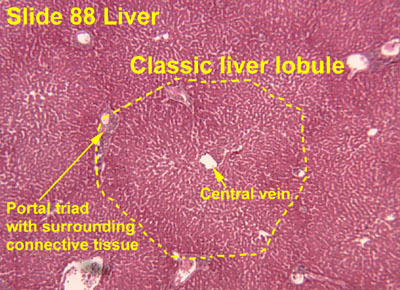
| classical lobule[2] | hexagonal; divided into concentric centrilobular, midzonal, periportal parts | anatomical |
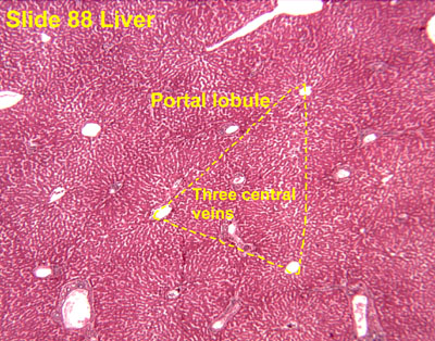
| portal lobule[3] | triangular; centered on a portal triad | bile secretion |
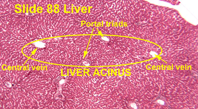
| acinus [4] | elliptical or diamond-shaped; divided into zone I (periportal), zone II (transition zone), and zone III (pericentral) | blood flow and metabolic |
The term "hepatic lobule", without qualification, typically refers to the classical lobule.
YouTube Encyclopedic
-
1/3Views:122 6702 90916 404
-
Hepatic lobule | Gastrointestinal system physiology | NCLEX-RN | Khan Academy
-
Liver 4, Lobules are amazing
-
Liver Architecture Made Simple! Classic Lobule, Hepatic Lobule, Heptaic Acinus
Transcription
So we've talked about a lot of vessels here. The main three that we should focus on are these guys right here, the portal vein, the hepatic artery and the common hepatic bile duct. Together these three make what's called the portal triad. It's one of the structures that are very important to identify in surgery and also is seen very commonly in the liver, when we look at smaller cuts of the organ. So if we were to focus in on a smaller section, like that. And blow it up so we can get a better picture of what's going on here. We can see that the liver itself, this giant fatty organ, is split up into smaller pieces or units, that are called hepatic lobules, hepatic lobules. And in the hepatic lobule we'll have a whole bunch of our hepatocytes or single liver cells, that are hanging out, sitting around here. Surrounding these hepatocytes is going to be a representation of our portal triad. That includes a branch that was part of the portal vein. A branch that was part of the hepatic artery and then a branch that will come together to make the common hepatic duct. So those three that make up your portal triad. Now this portal triad isn't found in just one part of the hepatic lobule. In fact, there's a bunch of them that surround the hepatocytes and make this very familiar shape, you might recall from chemistry. So it's this six-sided ring that makes up the hepatic lobule. That has six units of the portal triad surrounding the hepatocytes. And this is how we get nutrient rich blood to enter into the hepatic lobule, so that way these hepatocytes, these liver cells, can extract nutrients for the metabolism or the storage of macro molecules. And of course, hepatocytes are just like any other cell in the body, they need oxygen to survive. And so, oxygen will be carried in from our proper hepatic artery. So we'll supply the hepatocytes with oxygen, we'll supply them with nutrients from these branches off of the portal vein. They're called sinusoids, I'll write that off right here. Sinusoids that come off the portal vein and finally after nutrients have been extracted, oxygens been extracted and we need to send this blood back into the hepatic vein, all of the blood will collect at this very center right here, very conveniently called the central vein. So the central vein that sits in the very middle of our hepatic lobule, will take our nutrient and oxygen poor blood and then coalesce into a hepatic vein that'll be sent off to the heart for oxygenation by the lungs, and then passed by the intestines to receive nutrients from the absorption process we talked about. So that's how our liver works.
Structure
The hepatic lobule can be described in terms of metabolic "zones", describing the hepatic acinus (terminal acinus). Each zone is centered on the line connecting two portal triads and extends outwards to the two adjacent central veins. The periportal zone I is nearest to the entering vascular supply and receives the most oxygenated blood, making it least sensitive to ischemic injury while making it very susceptible to viral hepatitis. Conversely, the centrilobular zone III has the poorest oxygenation, and will be most affected during a time of ischemia.[5]
Portal triad
A portal triad (also known as portal canal, portal field[citation needed], portal area[citation needed], or portal tract[citation needed]) is a distinctive arrangement within lobules. It consists of the following five structures:[6]
- proper hepatic artery, an arteriole branch of the hepatic artery that supplies oxygen
- hepatic portal vein, a venule branch of the portal vein, with blood rich in nutrients but low in oxygen
- one or two small bile ductules of cuboidal epithelium, branches of the bile conducting system.
- lymphatic vessels
- branch of the vagus nerve
The misnomer "portal triad" traditionally has included only the first three structures, and was named before lymphatic vessels were discovered in the structure.[7][8] It can refer both to the largest branch of each of these vessels running inside the hepatoduodenal ligament, and to the smaller branches of these vessels inside the liver.
In the smaller portal triads, the four vessels lie in a network of connective tissue and are surrounded on all sides by hepatocytes. The ring of hepatocytes abutting the connective tissue of the triad is called the periportal limiting plate.
-
 Portal triad
 -
 Labeled sketch of a portal canal
 -
 Portal triad of a rat liver, 1 branch of hepatic artery, 2 branch of portal vein, 3 bile duct
 -
 Portal triad of mouse liver. 1= bile duct, 2= branch of hepatic artery, 3= branch of portal vein, 4= lymphatic vessels


Periportal space
The periportal space (Latin: spatium periportale), or periportal space of Mall,[9] is a space between the stroma of the portal canal and the outermost hepatocytes in the hepatic lobule, and is thought to be one of the sites where lymph originates in the liver.[10]
Fluid (residual blood plasma) that is not taken up by hepatocytes drains into the periportal space, and is taken up by the lymphatic vessels that accompany the other portal triad constituents.
Function

Zones differ by function:
- zone I hepatocytes are specialized for oxidative liver functions such as gluconeogenesis, β-oxidation of fatty acids and cholesterol synthesis
- zone III cells are more important for glycolysis, lipogenesis and cytochrome P-450-based drug detoxification.[11] This specialization is reflected histologically; the detoxifying zone III cells have the highest concentration of CYP2E1 and thus are most sensitive to NAPQI production in acetaminophen toxicity.[12]
Other zonal injury patterns include zone I deposition of hemosiderin in hemochromatosis and zone II necrosis in yellow fever.[11]
Clinical significance
Bridging fibrosis, a type of fibrosis seen in several types of liver injury, describes fibrosis from the central vein to the portal triad.[13]
See also
References
- ^ Cell and Tissue Structure at U. Va.
- ^ Histology image: 88_03 at the University of Oklahoma Health Sciences Center
- ^ Histology image: 88_09a at the University of Oklahoma Health Sciences Center
- ^ Histology image: 88_09b at the University of Oklahoma Health Sciences Center
- ^ B.R. Bacon; J.G. O'Grady; A.M. Di Bisceglie; J.R. Lake (2006). Comprehensive Clinical Hepatology. Elsevier Health Sciences. ISBN 0-323-03675-9.
- ^ Mescher, Anthony L. (2013). Junqueira's Basic Histology text and atlas. McGraw-Hill Education. p. 333. ISBN 978-0-07-180720-3.
- ^ Vander's Human Physiology, The Mechanisms of Body Function. 26 September 2016. ISBN 9781478436232.
- ^ "Physiology of the Hepatic Vascular System". www.vivo.colostate.edu. Retrieved 2018-12-04.
- ^ Roderick N. M. MacSween (2007). MacSween's pathology of the liver. Elsevier Health Sciences. pp. 44–. ISBN 978-0-443-10012-3. Retrieved 23 May 2011.
- ^ Ross, Michael H.; Pawlina, Wojciech (2006). Histology: A Text and Atlas. Lippincott Williams & Wilkins. p. 426. ISBN 0-7817-7221-4.
- ^ a b E.R. Schiff; M.F. Sorrell; W.C. Maddrey, eds. (2007). Schiff's Diseases of the Liver, Tenth Edition. Lippincott William & Wilkins. ISBN 978-0-7817-6040-9.
- ^ M.J. Burns; S.L. Friedman; A.M. Larson (2009). "Acetaminophen (paracetamol) poisoning in adults: Pathophysiology, presentation, and diagnosis". In D.S. Basow (ed.). UpToDate. Waltham, MA: UpToDate.
- ^ "The liver ~ Medical student education – Tissupath". tissupath.com.au. Retrieved 20 June 2018.