
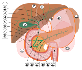
9. Gallbladder.
10–11. Right and left lobes of liver.
12. Spleen.
13. Esophagus.
14. Stomach.
15. Pancreas: 16. Accessory pancreatic duct, 17. Pancreatic duct.
18. Small intestine: 19. Duodenum, 20. Jejunum
21–22. Right and left kidneys.
The front border of the liver has been lifted up (brown arrow).[1]
Intrahepatic bile ducts compose the outflow system of exocrine bile product from the liver.
They can be divided into:[2]
- Lobar ducts (right and left hepatic ducts) - stratified columnar epithelium.
- Interlobar ducts (between the main hepatic ducts and the interlobular ducts) - pseudostratified columnar epithelium.
- Interlobular bile ducts (between the interlobar ducts and the lobules) - simple columnar epithelium.
- Intralobular bile ducts (cholangioles or Canals of Hering) - simple cuboidal epithelium, then by hepatocytes
- Bile canaliculi - two half-canaliculi formed by the hepatocytes facing the perisinusoidal space

YouTube Encyclopedic
-
1/3Views:8 12917 47346 262
-
Biliary tree | Root | Anatomy
-
Extrahepatic Biliary Apparatus | Cystohepatic Triangle - Anatomy Tutorial
-
Bile Ducts
Transcription
References
- ^ Standring S, Borley NR, eds. (2008). Gray's anatomy : the anatomical basis of clinical practice. Brown JL, Moore LA (40th ed.). London: Churchill Livingstone. pp. 1163, 1177, 1185–6. ISBN 978-0-8089-2371-8.
- ^ Roderick N. M. MacSween; Alastair D. Burt; Bernard Portmann; Linda D. Ferrell (2007). MacSween's pathology of the liver. Elsevier Health Sciences. p. 518. ISBN 978-0-443-10012-3.
This human digestive system article is a stub. You can help Wikipedia by expanding it. |