
| Trigeminal ganglion | |
|---|---|
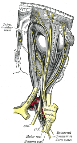 Nerves of the orbit. Seen from above. (Semilunar ganglion visible near bottom.) | |
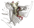
 Distribution of the maxillary and mandibular nerves, and the submaxillary ganglion. (Semilunar ganglion visible in upper left.) | |
| Details | |
| Identifiers | |
| Latin | ganglion trigeminale, ganglion semilunare (Gasseri) |
| MeSH | D012668 |
| TA98 | A14.2.01.014 |
| TA2 | 6194 |
| FMA | 52618 |
| Anatomical terms of neuroanatomy | |
The trigeminal ganglion (also known as: Gasserian ganglion, semilunar ganglion, or Gasser's ganglion) is the sensory ganglion of each trigeminal nerve (CN V). The trigeminal ganglion is located within the trigeminal cave (Meckel's cave), a cavity formed by dura mater.
YouTube Encyclopedic
-
1/5Views:16 456773 201554 339140 18110 109
-
Trigeminal Ganglion & nerve
-
Neurology | Trigeminal Nerve: Cranial Nerve V
-
Trigeminal Nerve Anatomy - Cranial Nerve 5 Course and Distribution
-
Anatomy Dissected: Cranial Nerve V (trigeminal nerve)
-
TRIGEMINAL GANGLION || ANATOMY || Meckel's cave || Trigeminal neuralgia ||
Transcription
Anatomy
The trigeminal ganglion contains cell bodies of the pseudo-unipolar sensory neurons of the trigeminal nerve which extend their axons both distally/peripherally into the three divisions of the trigeminal nerve on the one end, and proximally/centrally to the brainstem on the other end; the trigeminal root extends from the trigeminal ganglion to the ventrolateral aspect of the pons.[1]
The trigeminal ganglion is situated within the trigeminal cave (or Meckel's cave), a cerebrospinal fluid-filled cavity formed by a double layer[1] of dura mater overlying[1][2] the trigeminal impression near the apex of the petrous part of the temporal bone.[2]
Structure
The trigeminal ganglion is somewhat crescent-shaped, with its convexity directed anterolaterally. From its convex border arise the ophthalmic nerve (V1), maxillary nerve (V2), and mandibular nerve (V3).[2]
The ganglion receives, on its medial side, filaments from the carotid plexus of the sympathetic. It issues minute branches to the tentorium cerebelli, and the dura mater in the middle cranial fossa.[citation needed]
Relations
Medially to the trigeminal ganglion are the internal carotid artery, and the posterior part of the cavernous sinus.[2]
The motor root of the trigeminal nerve passes beneath the trigeminal ganglion to exit the skull through the foramen ovale.[2] The greater petrosal nerve passes underneath the trigeminal ganglion to reach the foramen lacerum.[3]: 498, 509
Clinical significance
Herpes virus dormancy
After recovery from a primary herpes infection, the virus is not cleared from the body, but rather lies dormant in a non-replicating state within the trigeminal ganglion.[4]
Lesions
If the trigeminal ganglion is damaged, by infection or surgery, it gives rise to the trigeminal trophic syndrome, which involves paresthesias and anesthesia, and may lead to erosions of the nasal ala.[citation needed]
Ablation in trigeminal neuralgia
The thermocoagulation or injection of glycerol into the trigeminal ganglion has been used in the treatment of trigeminal neuralgia.[citation needed]
Other animals
Rodents
In rodents, the trigeminal ganglion is important as it is the first part of the pathway from the whiskers to the brain. Cell bodies of the whisker primary afferents are found here. These afferents are mechanoreceptor cells that fire in response to whisker deflection.[citation needed]
There are around 26,000–43,000 cell bodies in rodent trigeminal ganglion. It is possible that there are two distinct (or perhaps continuous) populations of cells having slowly and rapidly adapting responses to stimuli.[citation needed]
It is found at the base of the skull and projects to trigeminal brain stem areas including principalis, spinal trigeminal nucleus, interpolaris, and caudalis.[citation needed]
Additional images
-
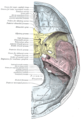 Base of the skull. Upper surface.
 -
 Nerves of the orbit, and the ciliary ganglion. Side view.
 -
 The otic ganglion and its branches.
 -
 Trigeminal ganglion
 -
 Trigeminal ganglion. Deep dissection. Superior view.


References
- ^ a b c Huff, Trevor; Weisbrod, Luke J.; Daly, Daniel T. (2022), "Neuroanatomy, Cranial Nerve 5 (Trigeminal)", StatPearls, Treasure Island (FL): StatPearls Publishing, PMID 29489263, retrieved 2023-01-03
- ^ a b c d e Gray, Henry (1918). Gray's Anatomy (20th ed.). p. 886.
- ^ Sinnatamby, Chummy S. (2011). Last's Anatomy (12th ed.). ISBN 978-0-7295-3752-0.
- ^ Verjans GM, Hintzen RQ, van Dun JM, et al. (2007). "Selective retention of herpes simplex virus-specific T cells in latently infected human trigeminal ganglia". Proc. Natl. Acad. Sci. U.S.A. 104 (9): 3496–501. Bibcode:2007PNAS..104.3496V. doi:10.1073/pnas.0610847104. PMC 1805572. PMID 17360672.
External links
- Diagram at University of Manitoba
- Diagram (as "Gasserian Ganglion") at frca.co.uk
- MedEd at Loyola grossanatomy/dissector/labs/h_n/cranium/cn3_1a.htm
- ancil-484 at NeuroNames
- cranialnerves at The Anatomy Lesson by Wesley Norman (Georgetown University) (V)
| National | |
|---|---|
| Other | |