
| Renal calyx | |
|---|---|
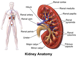 Kidney, with major and minor calyces labeled near bottom. | |
 An image showing just the pelvis and calices of the kidneys, with the rest of the kidney removed, from a dissected cow and seal specimen. These vary greatly in size and number depending on species.[citation needed] | |
| Details | |
| Precursor | Ureteric bud |
| System | Urinary system |
| Identifiers | |
| Latin | calices renales |
| MeSH | D007670 |
| FMA | 284558 |
| Anatomical terminology | |
The renal calyces are conduits in the kidney through which urine passes. The minor calyces form a cup-shaped drain around the apex of the renal pyramids. Urine formed in the kidney passes through a renal papilla at the apex into the minor calyx; 4-5 minor calyces converge to form a major calyx through which urine passes into the renal pelvis (which in turn drains urine out of the kidney through the ureter).[1]
YouTube Encyclopedic
-
1/3Views:58 94858 9941 352
-
Renal System 4, Paranchyma, calyces and pelvis.
-
Kidney anatomy
-
RENAL HILUM VARIANT VESSELS Atrophic Pelvi-Calyces and Clinical Significance – Sanjoy Sanyal
Transcription
Function
Peristalsis of the smooth muscle originating in pace-maker cells originating in the walls of the calyces propels urine through the renal pelvis and ureters to the bladder. The initiation is caused by the increase in volume that stretches the walls of the calyces. This causes them to fire impulses which stimulate rhythmical contraction and relaxation, called peristalsis. Parasympathetic innervation enhances the peristalsis while sympathetic innervation inhibits it.
Clinical significance

A "staghorn calculus" is a kidney stone that may extend into the renal calyces.
A renal diverticulum is diverticulum of renal calyces.[2][3]
See also
References
- ^ Martini, Frederic; Tallitsch, Robert B.; Nath, Judi L. (2017). Human Anatomy (9th ed.). Pearson. p. 689. ISBN 9780134320762.
- ^ Krzeski, T; Witeska, A; Borówka, A; Pypno, W (September 1981). "Diverticula of renal calyces". International Urology and Nephrology. 13 (3): 231–235. doi:10.1007/BF02082420. PMID 6799417. S2CID 275191.
- ^ Chong, TW; Bui, MH; Fuchs, GJ (Nov 2000). "Calyceal diverticula. Ureteroscopic management". The Urologic Clinics of North America. 27 (4): 647–54. doi:10.1016/s0094-0143(05)70114-2. PMID 11098763.
External links
- Anatomy photo:40:06-0108 at the SUNY Downstate Medical Center - "Posterior Abdominal Wall: Internal Structure of a Kidney"
- Anatomy photo:40:06-0109 at the SUNY Downstate Medical Center - "Posterior Abdominal Wall: Internal Structure of a Kidney"
- Histology image: 15901loa – Histology Learning System at Boston University - "Urinary System: neonatal kidney"
- posteriorabdomen at The Anatomy Lesson by Wesley Norman (Georgetown University) (renalpelvis)
- Diagram at bway.net