
| Bowman's capsule | |
|---|---|
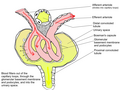
 Glomerulus (red), Bowman's capsule (blue) and proximal tubule (green) | |
| Details | |
| Precursor | Metanephric blastema |
| Location | Nephron of kidney |
| Identifiers | |
| Latin | capsula glomeruli |
| MeSH | D050476 |
| FMA | 15626 |
| Anatomical terminology | |
Bowman's capsule (or the Bowman capsule, capsula glomeruli, or glomerular capsule) is a cup-like sac at the beginning of the tubular component of a nephron in the mammalian kidney that performs the first step in the filtration of blood to form urine. A glomerulus is enclosed in the sac. Fluids from blood in the glomerulus are collected in the Bowman's capsule.
YouTube Encyclopedic
-
1/3Views:258 8941 67630 423
-
GLOMERULAR FILTRATION made easy!!
-
Function of Bowman's Capsule
-
Filtration in Renal Corpuscle (Glomerulus and Bowman's Capsule)
Transcription
hiya welcome to biomed sessions with me Ruz today we're going to be discussing Glomerular filtration here we have our renal corpuscle which is a structure essential for the filtration of blood in the nephrons of the kidney one part of the renal corpuscle is the glomerulus which is basically a network of capillaries the other part is the surrounding bowman's capsule most capillaries have an arterial end and a venous end this is not the case here as blood flows through the glomerulus from the afferent arteriole to the efferent arteriole and as this occurs, components of the blood are filtered out The fluid that enters the capsule is called glomerular filtrate and filtration occurs across or to filtration barrier and filtration occurs across an ultrafiltration barrier. A good analogy for the ultrafiltration barrier is a kitchen strainer. Now if you pour a nice herby broth of vegetables into the strainer you will see that large vegetables are left behind whereas water and anything that dissolves in it plus tiny particles are able to pass through. with our glomerulus, molecules less than one point eight nanometers are freely filtered out whereas molecules more than 3.6nm are not filtered. Let's take a look at a zoomed in section of the ultrafiltration barrier . As you can see there are three layers. Our bottom layer is the endothelium of the capillary which contains these pores known as fenestrations. This layer basically lets everything through except for blood cells. Our middle layer is the basement membrane which prevents the filtration of large proteins. And our outer layer consists of podocytes -- part of the bowman's capsule. These look like monsters wrapping their arms around the layers below and they themselves have many finger-like projections called pedicels are so close to each that there are just narrow filtration slits between them which allow only small molecules to pass through. One thing to note is that the ultrafiltration barrier As all 3 layers contain negatively charged glycoproteins it is difficult for negative molecules to pass through Hence serum albumin is not filtered (despite being in the size range). Ultrafiltration of blood to form glomerular filtrate depends on a balance between the forces that favor filtration and those that oppose it. In general we can refer to these forces as starling forces in order to fully understand glomerular filtration you need know about hydrostatic and oncotic pressure, so I'm going to give you a very simplified explanation. Hydrostatic pressure refers to the force a fluid exerts on the walls of it's compartment (this would be either the walls of the capillaries or the bowman's capsule). I like to think of it I like to think of it as 'pushing' because it is kinda like the way water pushes on the inside of a water balloon as it's being filled up but in this instance the fluid can be pushed out. Oncotic pressure is pressure exerted by plasma proteins on the walls of the compartment in which they are contained. It kinda has a sponge-like effect encouraging fluid to be drawn in, therefore I like to think of pressure as 'pulling'. The major driving force for filtration filtration is the hydrostatic pressure of the glomerulus which forces fluid out of the capillary. This is opposed by hydrostatic pressure of the bowman's capsule and the oncotic pressure of the glomerular capillary protein. [NOTE: [NOTE: we tend to ignore oncotic pressure of the bowman's capsule as only tiny amounts of protein are usually present in the glomerular filtrate] Our Net filtration pressure (NFP) equals to the pressures favouring filtration minus the pressures opposing filtration i.e. hydrostatic pressure of the glomerulus minus hydrostatic pressure of the bowman's capsule minus oncotic pressure of the glomerular capillary protein which is equal to ten millimeters of mercury There are many nephrons and hence there are many renal corpuscles in each kidney. Glomerular Filtration Rate GFR for short, is the total amount of filtrate formed by all the renal corpuscles in both kidneys per minute it can be used as a clue to assess whether an individual has kidney impairment. GFR not only takes into account NFP but also Surface area available for filtration and permeability of glomeruli . In fact, equals to the product of Surface area and permeability multiplied by NFP which can be condensed to the filtration coefficient multiplied by NFP. The permeability and surface area of glomerular capillaries tend to be greater compared to other capillaries due to the fenestrations and extensive branching and looping, hence the filtration coefficient is high and so there is a high degree of filtration in the glomerulus. It is important to note when considering GFR we must also take into account the number of functioning nephrons and how effectively the glomeruli filter blood. So How can GFR be changed? Well of course by altering either the filtration coefficient or NFP. For example if we constrict the afferent arteriole, the Hydrostatic Pressure of the glomerular blood will decrease due to the reduction of blood available for filtration. as this pressure is associated with NFP will also decrease and hence GFR will decrease. In a later video, we will go into more detail about the control of GFR and how it is estimated. But for now I would like you to understand that the glomerulus alongside the Bowman's capsule is highly specialized in the filtration of blood & the Glomerular filtration rate, GFR, is a good indicator of how well the kidneys are working. so if this tutorial was helpful to you show it! by liking the video & if you would like to see more tutorials from me -- do subscribe. Ok see you in another video! bye :)
Structure
Outside the capsule, there are two "poles":
- The vascular pole, polus vascularis is the side with the afferent arteriole and efferent arteriole.
- The urinary pole, polus urinarius is the side with the proximal convoluted tubule.
Inside the capsule, the layers are as follows, from outside to inside:[citation needed]
- Parietal layer—A single layer of simple squamous epithelium. Does not function in filtration.
- Bowman's space (or "urinary space", or "capsular space")—Between the visceral and parietal layers, into which the filtrate enters after passing through the filtration slits.[1]
- Visceral layer—Lies just above the thickened glomerular basement membrane and is made of podocytes. Beneath the visceral layer lie the glomerular capillaries.[citation needed]
- Filtration barrier—The filtration barrier is composed of the fenestrated endothelium of the glomerular capillaries, the fused basal lamina of the endothelial cells and podocytes, and the filtration slits of the podocytes. The barrier permits the passage of water, ions, and small molecules from the bloodstream into the Bowman's space. The barrier prevents the passage of large and/or negatively charged proteins (such as albumin). The basal lamina of the filtration barrier is composed of three layers. The first layer is the lamina rara externa, adjacent to the podocyte processes. The second layer is the lamina rara interna, adjacent to the endothelial cells. The final layer is the lamina densa which is a darker central zone of the basal lamina. It consists of the meshwork of type IV collagen and laminin which act as a selective macromolecular filter.[citation needed]


Function
The process of filtration of the blood in the Bowman's capsule is ultrafiltration (or glomerular filtration), and the normal rate of filtration is 125 ml/min, equivalent to 80 times the daily blood volume.[citation needed] It is a major site for blood filtration (including glomerulus)
Any proteins under roughly 30 kilodaltons can pass freely through the membrane, although there is some extra hindrance for negatively charged molecules due to the negative charge of the basement membrane and the podocytes.[citation needed]
Any small molecules such as water, glucose, salt (NaCl), amino acids, and urea pass freely into Bowman's space, but cells, platelets and large proteins do not.[citation needed]
As a result, the filtrate leaving the Bowman's capsule is very similar to blood plasma (filtrate or glomerular filtrate is composed of blood plasma minus plasma protein i.e. it contains all the components of blood plasma except the proteins) in composition as it passes into the proximal convoluted tubule.[citation needed]
Clinical significance

Measuring the glomerular filtration rate (GFR) is a diagnostic test of kidney function.[3]
A decreased GFR may be a sign of kidney failure.[citation needed]
A number of diseases can result in various problems within the glomerulus. Examples include acute proliferative (endocapillary) glomerulonephritis, mesangioproliferative glomerulonephritis, mesangiocapillary (membranoproliferative) glomerulonephritis, acute crescentic glomerulonephritis, focal segmental glomerulonephritis, and diabetic glomerulosclerosis.[citation needed]
History
Bowman's capsule is named after Sir William Bowman (1816–1892), a British surgeon and anatomist.[4] However, thorough microscopical anatomy of kidney including the nephronic capsule was first described by a Ukrainian surgeon and anatomist from the Russian Empire, Prof. Alexander Schumlansky (1748–1795), in his 1782 doctoral thesis "De structura renum" ("About Kidney Structure", in Latin); thus, much prior to Bowman.[5]
Together with the glomerulus it is known as a renal corpuscle, or a Malpighian corpuscle, named after Marcello Malpighi (1628–1694), an Italian physician and biologist. This name is not used widely anymore, probably to avoid confusion with Malpighian bodies of the spleen.[citation needed]
See also
- Mesangium
- Glomerulus (kidney)
- Blood–brain barrier
- List of distinct cell types in the adult human body
Additional images
-
 Glomerulus.

References
- ^ Histology image:22401lba from Vaughan, Deborah (2002). A Learning System in Histology: CD-ROM and Guide. Oxford University Press. ISBN 978-0195151732.
- ^ Table 4 in: Hodgin, Jeffrey B.; Bitzer, Markus; Wickman, Larysa; Afshinnia, Farsad; Wang, Su Q; O'Connor, Christopher; Yang, Yan; Meadowbrooke, Chrysta; Chowdhury, Mahboob; Kikuchi, Masao; Wiggins, Jocelyn E.; Wiggins, Roger C. (2015). "Glomerular Aging and Focal Global Glomerulosclerosis: A Podometric Perspective". Journal of the American Society of Nephrology. 26 (12): 3162–3178. doi:10.1681/ASN.2014080752. ISSN 1046-6673. PMC 4657829. PMID 26038526.
- ^ Romagnani, Paola; Anders, Hans-Joachim (2019). "Excretory System". In Brüne, Martin; Schiefenhövel, Wulf (eds.). Oxford Handbook of Evolutionary Medicine. Oxford University Press. ISBN 978-0198789666.
- ^ Bowman, William; Royal Society of London. Philosophical transactions, v. 32, p. 57-80, 1842 (1842). On the structure and use of the Malpighian bodies of the kidney: with observations on the circulation through that gland. London: Taylor. OCLC 7714131.
{{cite book}}: CS1 maint: multiple names: authors list (link) CS1 maint: numeric names: authors list (link) - ^ Schumlansky, Aleksander (1782). Dissertatio Inauguralis Anatomica De Structura Renum Quam Pro Licentia Summos In Medicina Honores Et Privilegia Doctoralia Legitime Obtinendi In Inclyta Argentoratensium Universitate Solenni Eruditorum Examini Submittit Alexander Schumlansky Poltawo-Russus Die XVI. Novembr. A. MDCCLXXXII (in Latin). Argentorati [Strasbourg]. p. 92.
External links
- Histology image: 16006loa – Histology Learning System at Boston University
- Diagram at ircc.edu