
| Ventromedial prefrontal cortex | |
|---|---|
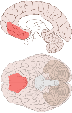 Ventromedial prefrontal cortex shown on medial and ventral views of the brain, reflecting approximate location of damage in patients with decision making deficits.[1] | |
 Medial surface of the brain with Brodmann's areas numbered. | |
| Details | |
| Identifiers | |
| Latin | cortex praefrontalis ventromedialis |
| Anatomical terms of neuroanatomy | |
The ventromedial prefrontal cortex (vmPFC) is a part of the prefrontal cortex in the mammalian brain. The ventral medial prefrontal is located in the frontal lobe at the bottom of the cerebral hemispheres and is implicated in the processing of risk and fear, as it is critical in the regulation of amygdala activity in humans.[2] It also plays a role in the inhibition of emotional responses, and in the process of decision-making and self-control. It is also involved in the cognitive evaluation of morality.
YouTube Encyclopedic
-
1/5Views:100 2533 61777 86916 370418
-
Emotions: cerebral hemispheres and prefrontal cortex | MCAT | Khan Academy
-
Functions of the Prefrontal Cortex
-
The Man With a Hole in His Brain
-
Personality Changes - Frontal lobe brain injury - TBI
-
Ventromedial.wmv
Transcription
So what we have here is a brain. And this is like we're above the person, looking down into their brain. To orient you, let me draw two eyes here. And this is the front of the brain and this is the back of the brain. And we're looking at it, like I said, from the top. Now, why are we looking at a brain? Well, I want to talk about a certain area of the brain known as the cerebral cortex and how it plays a role in emotions. And there's a number of different ways that you can divide up the cerebral cortex and organize it. So we're going to look at a few in terms of emotion. So one way that you can view the brain is in terms of hemispheres. And in your brain, you have two hemispheres. You have a left hemisphere and a right hemisphere. And the way you think of hemispheres, it's as if you draw the line down the brain. And you can split it into two different hemispheres. You have the left hemisphere and the right hemisphere. And there's actually some differences between the left and right hemisphere. Researchers actually found that positive emotions evoke more electrical activity on the left side of the brain than on the right. Whereas negative emotions tend to elicit more activity in the right hemisphere. Now, you might be wondering how did they figure that out? Well, what they did is they had participants in the research study watch a TV screen. So let's draw a TV here. And this was several years ago. So I bet TVs had bunny ears back then. So we'll draw some bunny ears here. So what this research study did was they had their participants watch films, either evoked pleasure or disgust. And pleasure films were things like the kind of videos that people share with each other on YouTube. I mean obviously YouTube back then didn't exist. So they just got films of things that evoked positive emotions, like puppies playing in flowers and actually a gorilla taking a bath at the zoo. I guess that does bring quite a bit of pleasure. But that wouldn't be the first thing that came to my mind. And they had them watch other videos that evoked feelings of disgust. And these were like shock films, like videos of third-degree burns and very like gory leg amputations, things that would evoke negative emotions. And while these participants were watching these films, the researchers videotaped their facial expressions and also recorded their brain activity through something called an EEG recording. And EEG recordings basically measure the electrical activity of your brain. And what they found was that pleasure films increase activity in the left hemisphere because the pleasure films are associated with positive emotions. Now, it's not to say that there wasn't any activity in the right hemisphere. There's just more activity in the left hemisphere because positive emotions increase activity in the left hemisphere. And the same could be said for the disgusting shock films. On EEG recording, they found that participants had more activity in the right hemisphere and these disgusting films are more associated with negative emotions, like fear and disgust. So I think it's pretty neat that while the brain is such a complicated structure, is that you can actually just split it down the middle and think of it in terms of left and right. So you can associate the left hemisphere with positive emotions and the right hemisphere with negative emotions. But that's not all in terms of hemispheres. This concept of left and right hemispheres becoming more active in certain situations can also be applied to social interaction and how outgoing and sociable you are. And in a different study, researchers observed a group of four-year-olds playing in a group. So I'm going to draw some little kids here. So you have someone here. The researchers watched the kids interacting in groups and saw how they interacted with each other. So here are two guys. And you can see them sharing in a cool toy here. And check out their expression. They're smiling and they look like they're having a great time. And some kids, that's how they act. Other kids tend to be more isolative. Some kids like to isolate a little bit more and be alone. And here's an example. Here's one kid here. He's just kind of sitting. He's all alone. He's frowning. He's kind of isolating over here. So after observing and noting how these children behaved, they did a similar experiment to what we mentioned above. And they took EEG recordings. And notice something kind of interesting. That the kids who were more sociable, playing in a group, they tended to have an increased level of activity in their left hemisphere. So I'll put "sociable." On the other hand, they noticed that the children who isolated more, like this guy here, they tended to have more activity in the right hemisphere. And to represent that, I'll write "isolative." And of course there's been other studies as well. And other research shows that people with more active left hemispheres, they tend to be more interested, joyful, and enthusiastic about things. Whereas those with more active right hemispheres, tend to be more timid, fearful, avoidant, and even depressed. So that's the basic overview of emotions in terms of brain hemispheres. Now another way you can look at the cerebral cortex is by dividing it into functional divisions. So I'm going to erase these structures up here. And some of these functional divisions you can already see in different colors here. But the one that I want to focus on is the prefrontal cortex. And the prefrontal cortex is basically this area right here, this area all the way in the front of your brain. And it's actually right behind your forehead. And this area of the brain is responsible for many high order functions, like language, information processing, all the things that you think of that make humans, humans-- the ability to solve math problems, think through philosophical issues. Sometimes these are referred to as very like cerebral activities. And where do you think that term comes from? Well, the cerebral cortex and the prefrontal cortex, a part of the cerebral cortex. And like I said, the prefrontal cortex is really what distinguishes humans. And because of that, it's extremely well developed in humans. And it undergoes the greatest amount of development after birth. I mean think of how you become more mature as you get older, or so they say. In terms of the prefrontal cortex, you use this part of the brain when you're trying to solve problems, make decisions, and manage how you behave in social situations. For instance, you probably behave differently during a job interview than you do during a wild sporting event. And even if you don't, you know that there are certain norms and expectations in job interviews than there are in sporting events. And the ability to know that comes from your prefrontal cortex. And I mention this because in another video, we talked about the amygdala. And the amygdala causes fear, anxiety, anger, and aggression. And I'm sure you've had the experience in your life where you felt very angry at someone. And when you have those sorts of feelings, maybe your primal reaction is to attack the person or physically hit them. But of course, many people don't do that. And why is that? Well, because they have a well-developed prefrontal cortex and the ability to understand, well, violence isn't the answer. You shouldn't attack someone, no matter how you feel. And that thinking that you have your mind where you say no, no, I should restrict my behavior, I should walk away, all those thoughts that you have come from the prefrontal cortex because that helps to manage how you behave in social situations. One question that's interesting to consider is what would happen if you damaged your prefrontal cortex? How would you act? Well, there's a famous example of this where someone actually did have that happen to them. And his name was Phineas Gage. Now, Phineas Gage was 25 years old, back when it was 1848. And he was a railroad worker. So I'll draw some train tracks here. And as part of building this railroad, they had to do controlled explosions to make way for it. And Phineas Gage was responsible for overseeing that. So here he is. And he was just overseeing these explosions. Now during one of these explosions, an iron rod was sent flying through the air. And it actually penetrated through his skull. It went in one side and out the other. And during its course through his skull, it destroyed much of his prefrontal cortex. The man that Phineas Gage was after the accident was not the same man he was before. You see before, his friends and coworkers known as a hard working and well-liked guy. After he experienced this trauma to his head, he became rude and gruff. He swore a lot and just behaved inappropriately overall. And that's because he no longer had a functioning prefrontal cortex. So that's the cerebral cortex. So if you ever wonder what makes a human, human, well, a lot of it's actually right behind your forehead, in the area known as the prefrontal cortex.
Anatomy
While the ventromedial prefrontal cortex does not have a universally agreed on demarcation, in most sources, it is equivalent to the ventromedial reward network of Öngür and Price.[3] This network includes Brodmann area 10, Brodmann area 14, Brodmann area 25, and Brodmann area 32, as well as portions of Brodmann area 11, Brodmann area 12, and Brodmann area 13.[4] However, not all sources agree on the boundaries of the area. Different researchers use the term ventromedial prefrontal cortex differently. Sometimes, the term is saved for the area above the medial orbitofrontal cortex, while at other times, 'ventromedial prefrontal cortex' is used to describe a broad area in the lower (ventral) central (medial) region of the prefrontal cortex, of which the medial orbitofrontal cortex constitutes the lowermost part. This latter, broader area, corresponds to the area damaged in patients with decision-making impairments investigated by António Damásio and colleagues (see diagram, and below).
The ventromedial prefrontal cortex is connected to and receives input from the ventral tegmental area, amygdala, the temporal lobe, the olfactory system, and the dorsomedial thalamus. It, in turn, sends signals to many different brain regions including; The temporal lobe, amygdala, the lateral hypothalamus, the hippocampal formation, the cingulate cortex, and certain other regions of the prefrontal cortex.[5] This huge network of connections affords the vmPFC the ability to receive and monitor large amounts of sensory data and to affect and influence a plethora of other brain regions, particularly the amygdala.
Function
Functional differences between the orbitofrontal and ventromedial areas of the pre-frontal cortex have not yet been clearly established, although the areas of the ventromedial cortex superior to the orbitofrontal cortex are much less associated with social functions and more with pure emotion regulation. Research in developmental neuroscience also suggested that neural networks in the ventromedial prefrontal cortex are rapidly developing during adolescence and young adulthood supporting emotion regulation through the amygdala,[6] being associated with a decrease in cortisol levels.
There are only a few reports of people with early-onset vmPFC damage during childhood, but these individuals tend to have severe antisocial behavior and impaired moral judgment. Compared to individuals with damage later in life, their behavior pattern is similar but more severe.[7] It is also considered central to the physiology of anxiety and mood disorders. However, the precise mechanisms by which vmPFC contributes to affective processing are not fully understood.[2]
Decision making
Patients with bilateral lesions of the vmPFC develop severe impairments in personal and social decision-making[5][8] even though most of their intellectual ability is preserved.[8][9] For instance, they have difficulties in choosing between options with uncertain outcomes, whether the uncertainty is in the form of a risk or of an ambiguity.[10] After their lesion, these patients have an impaired capacity to learn from their mistakes, making the same decisions again and again even though they lead to negative consequences. These patients choose alternatives that give immediate rewards, but seem to be blind to the future consequences of their actions.[8] However, the underlying mechanisms of this behavior are not yet fully understood.[8]
Damage to the ventromedial prefrontal cortex (especially in the right hemisphere) has been connected with deficits in detecting irony, sarcasm, and deception.[11] Subjects with damage in this area have been found to be more easily influenced by misleading advertising.[12] This has been attributed to a disruption of a "false tagging mechanism" which provides doubt and skepticism of new beliefs.
People with damage to the ventromedial prefrontal cortex still retain the ability to consciously make moral judgments without error, but only in hypothetical situations presented to them. They are severely impaired in making personal and social decisions.[13] There is a gap in reasoning when applying the same moral principles to similar situations in their own lives. The result is that people make decisions that are inconsistent with their self professed moral values.[5] People with early damage to the ventromedial prefrontal cortex are more likely to endorse self-serving actions that break moral rules or cause harm to others. This is especially true for patients whose damage occurred the earliest in life.[14]
Emotions and an understanding of social norms are used to provide reasoning of the moral nature on our behaviors, beliefs, and the people around us. The vmPFC works as the neural basis in allowing emotion to influence moral judgement. In functional imaging studies, increased activity in the vmPFC is associated with thinking of these personal moral situations, while making harmless decisions does not.[15] Patients with vmPFC lesions made the same decision in impersonal and personal dilemmas. Dysfunction of the vmPFC causes failure in using correct moral emotion, which explains why these patients showed less emotional responses when facing these dilemmas.[16]
Regulation of emotion
The vmPFC plays an important role in regulating and inhibiting our response to emotions. VmPFC seems to use our emotional reactions to model our behavior and control emotional reactions in certain social situations. The inputs of the vmPFC provide it with information from the environment and the plans of the frontal lobe, and its outputs allow the vmPFC to control different physiological responses and behaviors. The role of the vmPFC is especially highlighted in people with damage to this region. A damaged vmPFC causes impairments of behavioral control and decision making, consequences which are rooted in emotional dysregulation.
The first and most famous case of someone with defects to this region was Phineas Gage, a railroad construction foreman who had his vmPFC bilaterally destroyed in an accident in 1848. Before his accident, Gage was described as “serious, industrious and energetic. Afterward he became childish, irresponsible, and thoughtless of others.”[17] Another patient with vmPFC damage wasted away his life savings on foolish investments and failed to make appropriate decisions in his personal life. In patients with vmPFC damage, evidence shows that there is a correlation between emotional disregulation and dysfunction in real world competencies.[17]
The amygdala plays a significant role in instigating the emotional reactions associated with anger and violence. With the vmPFC’s outputs to the amygdala, the vmPFC plays a part in preventing such behavior. Evidence has shown that impulsive murderers have decreased activity in the prefrontal cortex and increased activity in subcortical areas such as the amygdala. This imbalance can enhance actions that are created by negative emotions and limit the ability of the prefrontal cortex to control these emotions. Lower activation in the prefrontal cortex is also correlated with antisocial behavior. The dysfunction of the ventromedial cortex seems to, in part, be caused by lower levels of serotonin release.[17]
The vmPFC also is involved in courage. In experiments with participants allowing snakes to come near or away from them, acts of courage correlated with activation in the vmPFC, specifically the subgenual anterior cingulate cortex.[17][18]
Activation of the vmPFC is associated with successful suppression of emotional responses to a negative emotional signal.[19] Patients with vmPFC lesions show defects both in emotional response and emotion regulation.[9] Their emotional responsiveness is generally diminished and they show markedly reduced social emotions such as compassion, shame and guilt. These are emotions that are closely associated with moral values.[9] Patients also exhibit poorly regulated anger and frustration tolerance in certain circumstances.[9]
Patients with focal lesions in the vmPFC show personality changes such as lack of empathy, irresponsibility, and poor decision making. These traits are similar to psychopathic personality traits.[20] In addition, a correlation between individuals with a history of physical violence and decreased grey matter density in the vmPFC has been evidenced.[21] Lesions to the vmPFC are associated with resistance to depression, whereas lesions to the dorsolateral PFC is associated with vulnerability to depression.[22]
The right half of the ventromedial prefrontal cortex was associated with regulating the interaction of cognition and affect in the production of empathic responses. Hedonic (pleasure) responses were also associations to orbitofrontal cortex activity level by Morten Kringelbach. This finding contributes findings suggesting ventromedial prefrontal cortex being associated with preference judgement, possibly assigning the ventromedial prefrontal cortex a key role in constructing one's self. fMRI scans have found that the vmPFC is active when people think about themselves. There are cultural differences in the use of this region based on cultural differences in self-perception. Chinese subjects who think of the self in relation to the community have been found to utilize the vmPFC when thinking about their mothers, whereas American subjects do not.[23]
Studies with post-traumatic stress disorder (PTSD) also supported the idea that the ventromedial prefrontal cortex is an important component for reactivating past emotional associations and events, therefore essentially mediating pathogenesis of PTSD.[24][25] Dysfunction of the vmPFC has also been identified as playing a role in PTSD-affected parents' response to their own children's mental states.[26] Treatments geared to the activation of the ventromedial prefrontal cortex were therefore suggested for individuals and parent-child relationships affected by PTSD. The right half of the ventrolateral prefrontal cortex, being active during emotion regulation, was activated when participants were offered an unfair offer in a scenario. Specific deficits in reversal learning and decision-making have led to the hypothesis that the ventromedial prefrontal cortex is a major locus of dysfunction in the mild stages of the behavioral variant of frontotemporal dementia.[27] A study of patients with lesions in the right vmPFC showed a loss of empathy and theory of mind, showing that the brain regions is directly involved in empathy and mentalizing.[28]
The capacity for mature defense mechanisms such as intellectualization, compensation, reaction formation, and isolation has been tied to proper functioning of the right ventromedial prefrontal cortex, while more primitive defense mechanisms such as projection, splitting, verbal denial, and fantasy have been found to rely on other regions, primarily in the left hemisphere.[29]
Somatic marker hypothesis
One particularly notable theory of vmPFC function is the somatic marker hypothesis, accredited to António Damásio. By this hypothesis, the vmPFC has a central role in adapting somatic markers—emotional associations, or associations between mental objects and visceral (bodily) feedback—for use in natural decision making. This account also gives the vmPFC a role in moderating emotions and emotional reactions because whether the vmPFC decides the markers are positive or negative affects the appropriate response in a particular situation.[citation needed] However, a critical review of this hypothesis concluded that there is a need for additional empirical data to support the somatic marker theory.[19]
Extinction
Another role that the vmPFC plays is in the process of extinction, the gradual weakening and eventual cessation of a conditioned response, as studies have shown increased activation of the vmPFC after extinction training.[30] The specific role played by the vmPFC concerning extinction is not well understood, but it is believed that it plays a necessary role in the recall of extinction learning after a long period of time. Studies show that it aids in the consolidation of extinction learning.[31] A separate study has implicated the correlation between the cortical thickness of the vmPFC and the degree of extinction memory. Patients with larger vmPFCs tended to have lower responses to the extinct conditioned stimulus, therefore suggesting a superior extinction memory.[32] In general, the ventromedial prefrontal cortex plays a major role in the later stages of memory consolidation.[33]
Gender specific social cues
Ventromedial prefrontal cortex lesions were also associated with a deficit in processing gender specific social cues. One experiment tested the ability of patients with vmPFC lesions to categorize gender-specific names, attributes, and attitudes compared to patients with dorsolateral prefrontal cortex lesions and control subjects. Whereas the patients with dorsolateral prefrontal cortex lesions performed similarly to the control subjects on tests indicating gender stereotypes, patients with ventromedial prefrontal cortex lesions demonstrated impaired stereotypic social knowledge.[34]
Cocaine abuse
Frequent cocaine users have been shown to have lower than normal activity in the ventromedial prefrontal cortex. When asked to perform certain tasks that rely heavily on activation of this area of the brain, the cocaine users perform worse and have less prefrontal cortex activation than the control subjects.[35] The quantity of cocaine used was found to be inversely proportional to the level of activation.[36]
The prefrontal cortex is also physically affected by cocaine use. Chronic use has been shown to lead to a decrease in the amount of gray matter in the ventromedial prefrontal cortex. The decrease in gray matter and effect on behavior is analogous to a person having lesions throughout their medial prefrontal cortex.[35] Specifically, the pyramidal cells of the ventromedial prefrontal cortex are known to be linked with drug seeking behaviors.[37] Both an increased and decreased level of activity in these pyramidal cells has shown to lead to extinction of cocaine-seeking behaviors depending on when the activation takes place. Inactivation of these cells was needed to inhibit cocaine-seeking behavior after a longer duration of time, whereas activation was required to reduce the behavior soon after using cocaine.[38]
References
- ^ Bechara A, Damasio H, Tranel D, Anderson SW (January 1998). "Dissociation Of working memory from decision making within the human prefrontal cortex". The Journal of Neuroscience. 18 (1): 428–37. doi:10.1523/JNEUROSCI.18-01-00428.1998. PMC 6793407. PMID 9412519.
- ^ a b Motzkin JC, Philippi CL, Wolf RC, Baskaya MK, Koenigs M (February 2015). "Ventromedial prefrontal cortex is critical for the regulation of amygdala activity in humans". Biological Psychiatry. 77 (3): 276–284. doi:10.1016/j.biopsych.2014.02.014. PMC 4145052. PMID 24673881.
- ^ Ongür D, Price JL (March 2000). "The organization of networks within the orbital and medial prefrontal cortex of rats, monkeys and humans". Cerebral Cortex. 10 (3): 206–19. doi:10.1093/cercor/10.3.206. PMID 10731217.
- ^ Finger EC, Marsh AA, Mitchell DG, Reid ME, Sims C, Budhani S, Kosson DS, Chen G, Towbin KE, Leibenluft E, Pine DS, Blair JR (May 2008). "Abnormal ventromedial prefrontal cortex function in children with psychopathic traits during reversal learning". Archives of General Psychiatry. 65 (5): 586–94. doi:10.1001/archpsyc.65.5.586. PMC 3104600. PMID 18458210.
- ^ a b c Carlson NR (2013). Physiology of Behavior (11th ed.). Boston: Pearson.
- ^ Decety J, Michalska KJ (November 2010). "Neurodevelopmental changes in the circuits underlying empathy and sympathy from childhood to adulthood". Developmental Science. 13 (6): 886–99. doi:10.1111/j.1467-7687.2009.00940.x. PMID 20977559. S2CID 10647101.
- ^ Boes AD, Grafft AH, Joshi C, Chuang NA, Nopoulos P, Anderson SW (December 2011). "Behavioral effects of congenital ventromedial prefrontal cortex malformation". BMC Neurology. 11 (151): 151. doi:10.1186/1471-2377-11-151. PMC 3265436. PMID 22136635.
- ^ a b c d Bechara A, Tranel D, Damasio H (November 2000). "Characterization of the decision-making deficit of patients with ventromedial prefrontal cortex lesions". Brain. 123 ( Pt 11) (11): 2189–202. doi:10.1093/brain/123.11.2189. PMID 11050020.
- ^ a b c d Koenigs M, Young L, Adolphs R, Tranel D, Cushman F, Hauser M, Damasio A (April 2007). "Damage to the prefrontal cortex increases utilitarian moral judgements". Nature. 446 (7138): 908–11. Bibcode:2007Natur.446..908K. doi:10.1038/nature05631. PMC 2244801. PMID 17377536.
- ^ Fellows LK, Farah MJ (November 2007). "The role of ventromedial prefrontal cortex in decision making: judgment under uncertainty or judgment per se?". Cerebral Cortex. 17 (11): 2669–74. doi:10.1093/cercor/bhl176. PMID 17259643.
- ^ Zald DH, Andreotti C (October 2010). "Neuropsychological assessment of the orbital and ventromedial prefrontal cortex". Neuropsychologia. 48 (12): 3377–91. doi:10.1016/j.neuropsychologia.2010.08.012. PMID 20728457. S2CID 18918430.
- ^ Asp E, Manzel K, Koestner B, Cole CA, Denburg NL, Tranel D (2012). "A neuropsychological test of belief and doubt: damage to ventromedial prefrontal cortex increases credulity for misleading advertising". Frontiers in Neuroscience. 6: 100. doi:10.3389/fnins.2012.00100. PMC 3391647. PMID 22787439.
- ^ Bechara A, Tranel D, Damasio H (November 2000). "Characterization of the decision-making deficit of patients with ventromedial prefrontal cortex lesions". Brain. 123 (11): 2189–202. doi:10.1093/brain/123.11.2189. PMID 11050020.
- ^ Taber-Thomas BC, Asp EW, Koenigs M, Sutterer M, Anderson SW, Tranel D (April 2014). "Arrested development: early prefrontal lesions impair the maturation of moral judgement". Brain. 137 (Pt 4): 1254–61. doi:10.1093/brain/awt377. PMC 3959552. PMID 24519974.
- ^ Nicolle A, Goel V (2013). "What is the role of ventromedial prefrontal cortex in emotional influences on reason?". In Blanchette I (ed.). Emotion and Reasoning. Psychology Press.
- ^ Hu C, Jiang X (2014). "An emotion regulation role of ventromedial prefrontal cortex in moral judgment". Frontiers in Human Neuroscience. 8: 873. doi:10.3389/fnhum.2014.00873. PMC 4211379. PMID 25389402.
- ^ a b c d Carlson, N. (2012). Physiology of Behavior (11th ed.). Harlow: Prentice Hall.
- ^ Nili U, Goldberg H, Weizman A, Dudai Y (June 2010). "Fear thou not: activity of frontal and temporal circuits in moments of real-life courage". Neuron. 66 (6): 949–62. doi:10.1016/j.neuron.2010.06.009. PMID 20620879. S2CID 13941370.
- ^ a b Hänsel A, von Känel R (November 2008). "The ventro-medial prefrontal cortex: a major link between the autonomic nervous system, regulation of emotion, and stress reactivity?". BioPsychoSocial Medicine. 2 (21): 21. doi:10.1186/1751-0759-2-21. PMC 2590602. PMID 18986513.
- ^ Motzkin JC, Newman JP, Kiehl KA, Koenigs M (November 2011). "Reduced prefrontal connectivity in psychopathy". The Journal of Neuroscience. 31 (48): 17348–57. doi:10.1523/jneurosci.4215-11.2011. PMC 3311922. PMID 22131397.
- ^ Chester DS, Lynam DR, Milich R, DeWall CN (December 2017). "Physical aggressiveness and gray matter deficits in ventromedial prefrontal cortex". Cortex; A Journal Devoted to the Study of the Nervous System and Behavior. 97 (Supplement C): 17–22. doi:10.1016/j.cortex.2017.09.024. PMC 5716918. PMID 29073459.
- ^ Koenigs, Michael; Huey, Huey D; Calamia, Matthew; Raymont, Vanessa; Tranel, Daniel; Grafman, Jordan (2008). "Distinct regions of prefrontal cortex mediate resistance and vulnerability to depression". J Neurosci. 28 (47): 12341–12348. doi:10.1523/JNEUROSCI.2324-08.2008. PMC 2644261. PMID 19020027.
- ^ Zhu, Ying; Zhang, Li; Fan, Jin; Han, Shihui (2007). "Neural basis of cultural influence on self-representation". NeuroImage. 34 (3). Elsevier: 1310–1316. doi:10.1016/j.neuroimage.2006.08.047. ISSN 1053-8119. PMID 17134915. S2CID 11613104.
- ^ Insel TR (April 2009). "Disruptive insights in psychiatry: transforming a clinical discipline". The Journal of Clinical Investigation. 119 (4): 700–5. doi:10.1172/jci38832. PMC 2662575. PMID 19339761.
- ^ Koenigs M, Grafman J (October 2009). "Posttraumatic stress disorder: the role of medial prefrontal cortex and amygdala". The Neuroscientist. 15 (5): 540–8. doi:10.1177/1073858409333072. PMC 2771687. PMID 19359671.
- ^ Schechter DS, Moser DA, Giacobino A, Stenz L, Gex-Fabry M, Adouan W, Cordero MI, Suardi F, Manini A, Sancho-Rossignol A, Merminod G, Aue T, Ansermet F, Dayer AG, Rusconi-Serpa S. (epub May 29, 2015) Methylation of NR3C1 is related to maternal PTSD, parenting stress and maternal medial prefrontal cortical activity in response to child separation among mothers with histories of violence exposure. Frontiers in Psychology. http://journal.frontiersin.org/article/10.3389/fpsyg.2015.00690/abstract
- ^ Grossman M, Eslinger PJ, Troiani V, Anderson C, Avants B, Gee JC, McMillan C, Massimo L, Khan A, Antani S (October 2010). "The role of ventral medial prefrontal cortex in social decisions: converging evidence from fMRI and frontotemporal lobar degeneration". Neuropsychologia. 48 (12): 3505–12. doi:10.1016/j.neuropsychologia.2010.07.036. PMC 2949451. PMID 20691197.
- ^ Shamay-Tsoory SG, Tomer R, Berger BD, Aharon-Peretz J (April 2003). "Characterization of empathy deficits following prefrontal brain damage: the role of the right ventromedial prefrontal cortex". Journal of Cognitive Neuroscience. 15 (3): 324–37. doi:10.1162/089892903321593063. PMID 12729486. S2CID 8416412.
- ^ Northoff G (2010). "Region-based approach versus mechanism-based approach to the brain". Neuropsychoanalysis. 12 (2): 167–170. doi:10.1080/15294145.2010.10773640. S2CID 741542.
- ^ Madsen HB, Guerin AA, Kim JH (November 2017). "Investigating the role of dopamine receptor- and parvalbumin-expressing cells in extinction of conditioned fear". Neurobiology of Learning and Memory. 145: 7–17. doi:10.1016/j.nlm.2017.08.009. PMID 28842281. S2CID 26875742.
- ^ Quirk GJ, Russo GK, Barron JL, Lebron K (August 2000). "The role of ventromedial prefrontal cortex in the recovery of extinguished fear". The Journal of Neuroscience. 20 (16): 6225–31. doi:10.1523/JNEUROSCI.20-16-06225.2000. PMC 6772571. PMID 10934272.
- ^ Milad MR, Quinn BT, Pitman RK, Orr SP, Fischl B, Rauch SL (July 2005). "Thickness of ventromedial prefrontal cortex in humans is correlated with extinction memory". Proceedings of the National Academy of Sciences of the United States of America. 102 (30): 10706–11. Bibcode:2005PNAS..10210706M. doi:10.1073/pnas.0502441102. PMC 1180773. PMID 16024728. These results are a possible factor for explaining why different people show different degrees of controlling their fear.
- ^ Nieuwenhuis IL, Takashima A (April 2011). "The role of the ventromedial prefrontal cortex in memory consolidation". Behavioural Brain Research. 218 (2): 325–34. doi:10.1016/j.bbr.2010.12.009. hdl:2066/99828. PMID 21147169. S2CID 15659237.
- ^ Milne E, Grafman J (June 2001). "Ventromedial prefrontal cortex lesions in humans eliminate implicit gender stereotyping". The Journal of Neuroscience. 21 (12): RC150. doi:10.1523/JNEUROSCI.21-12-j0001.2001. PMC 6762729. PMID 11404442.
- ^ a b Carlson N (1977). Physiology of Behavior (11th ed.). Boston: Allyn and Bacon. pp. 621–622. ISBN 978-0-205-05706-1.
- ^ Bolla K, Ernst M, Kiehl K, Mouratidis M, Eldreth D, Contoreggi C, Matochik J, Kurian V, Cadet J, Kimes A, Funderburk F, London E (2004). "Prefrontal cortical dysfunction in abstinent cocaine abusers". The Journal of Neuropsychiatry and Clinical Neurosciences. 16 (4): 456–64. doi:10.1176/appi.neuropsych.16.4.456. PMC 2771441. PMID 15616172.
- ^ Kalivas PW, Volkow N, Seamans J (March 2005). "Unmanageable motivation in addiction: a pathology in prefrontal-accumbens glutamate transmission". Neuron. 45 (5): 647–50. doi:10.1016/j.neuron.2005.02.005. PMID 15748840. S2CID 2803383.
- ^ Van den Oever MC, Rotaru DC, Heinsbroek JA, Gouwenberg Y, Deisseroth K, Stuber GD, Mansvelder HD, Smit AB (November 2013). "Ventromedial prefrontal cortex pyramidal cells have a temporal dynamic role in recall and extinction of cocaine-associated memory". The Journal of Neuroscience. 33 (46): 18225–33. doi:10.1523/JNEUROSCI.2412-13.2013. PMC 3828471. PMID 24227731.
Further reading
- Bechara A, Tranel D, Damasio H (November 2000). "Characterization of the decision-making deficit of patients with ventromedial prefrontal cortex lesions". Brain. 123 ( Pt 11) (Pt 11): 2189–202. doi:10.1093/brain/123.11.2189. PMID 11050020.
- Quirk GJ, Russo GK, Barron JL, Lebron K (August 2000). "The role of ventromedial prefrontal cortex in the recovery of extinguished fear". The Journal of Neuroscience. 20 (16): 6225–31. doi:10.1523/JNEUROSCI.20-16-06225.2000. PMC 6772571. PMID 10934272.
- Hotz RL (2007-05-11). "Scientists Draw Link Between Morality And Brain's Wiring". Wall Street Journal.
- Carey B (2007-03-22). "Brain Injury Said to Affect Moral Choices". New York Times.
- Shamay-Tsoory SG, Tomer R, Berger BD, Aharon-Peretz J (April 2003). "Characterization of empathy deficits following prefrontal brain damage: the role of the right ventromedial prefrontal cortex". Journal of Cognitive Neuroscience. 15 (3): 324–37. doi:10.1162/089892903321593063. PMID 12729486. S2CID 8416412.
- Hooper CJ, Luciana M, Conklin HM, Yarger RS (November 2004). "Adolescents' performance on the Iowa Gambling Task: implications for the development of decision making and ventromedial prefrontal cortex". Developmental Psychology. 40 (6): 1148–58. doi:10.1037/0012-1649.40.6.1148. PMID 15535763.
- Elliott R, Rees G, Dolan RJ (April 1999). "Ventromedial prefrontal cortex mediates guessing". Neuropsychologia. 37 (4): 403–11. doi:10.1016/S0028-3932(98)00107-9. hdl:21.11116/0000-0001-A245-A. PMID 10215087. S2CID 18058688.
- Rahman S, Sahakian BJ, Hodges JR, Rogers RD, Robbins TW (August 1999). "Specific cognitive deficits in mild frontal variant frontotemporal dementia". Brain. 122 ( Pt 8) (8): 1469–93. doi:10.1093/brain/122.8.1469. PMID 10430832.
- Mehler J, Franck S (1995). Cognition on cognition. MIT Press. pp. 3–. ISBN 978-0-262-63167-9.
Intensity to Future Consequences Following Damage to Human Prefrontal Cortex
- Urry HL, van Reekum CM, Johnstone T, Kalin NH, Thurow ME, Schaefer HS, Jackson CA, Frye CJ, Greischar LL, Alexander AL, Davidson RJ (April 2006). "Amygdala and ventromedial prefrontal cortex are inversely coupled during regulation of negative affect and predict the diurnal pattern of cortisol secretion among older adults". The Journal of Neuroscience. 26 (16): 4415–25. doi:10.1523/JNEUROSCI.3215-05.2006. PMC 6673990. PMID 16624961.
- Paulus MP, Frank LR (July 2003). "Ventromedial prefrontal cortex activation is critical for preference judgments". NeuroReport. 14 (10): 1311–5. doi:10.1097/01.wnr.0000078543.07662.02. PMID 12876463. S2CID 5668392.
- Zald DH, Mattson DL, Pardo JV (February 2002). "Brain activity in ventromedial prefrontal cortex correlates with individual differences in negative affect". Proceedings of the National Academy of Sciences of the United States of America. 99 (4): 2450–4. Bibcode:2002PNAS...99.2450Z. doi:10.1073/pnas.042457199. PMC 122385. PMID 11842195.
- Tabibnia G, Satpute AB, Lieberman MD (April 2008). "The sunny side of fairness: preference for fairness activates reward circuitry (and disregarding unfairness activates self-control circuitry)". Psychological Science. 19 (4): 339–47. doi:10.1111/j.1467-9280.2008.02091.x. PMID 18399886. S2CID 15454802.
- Milne E, Grafman J (June 2001). "Ventromedial prefrontal cortex lesions in humans eliminate implicit gender stereotyping". The Journal of Neuroscience. 21 (12): RC150. doi:10.1523/JNEUROSCI.21-12-j0001.2001. PMC 6762729. PMID 11404442.
- Koenigs M, Huey ED, Raymont V, Cheon B, Solomon J, Wassermann EM, Grafman J (February 2008). "Focal brain damage protects against post-traumatic stress disorder in combat veterans". Nature Neuroscience. 11 (2): 232–7. doi:10.1038/nn2032. PMC 2693133. PMID 18157125.