
| Sphenoidal process of palatine bone | |
|---|---|
 Left palatine bone. Nasal aspect. Enlarged. | |
 Left palatine bone. Posterior aspect. Enlarged. | |
| Details | |
| Identifiers | |
| Latin | processus sphenoidalis |
| TA98 | A02.1.13.012 |
| TA2 | 810 |
| FMA | 59146 |
| Anatomical terms of bone | |
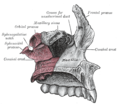
The sphenoidal process of palatine bone is a thin, superomedially directed plate of bone. It is smaller and more inferior compared to the orbital process of palatine bone.[1]
YouTube Encyclopedic
-
1/3Views:80542 36750 246
-
PALATINE BONE
-
Osteology of the Skull: 11 Palatine Bone
-
Maxilla and Palatine Bone.mov
Transcription
Anatomy
Surfaces
- The superior surface articulates with the root of the pterygoid process and the under surface of the sphenoidal concha, its medial border reaching as far as the ala of the vomer; it presents a groove which contributes to the formation of the pharyngeal canal.
- The medial surface is concave, and forms part of the lateral wall of the nasal cavity.
- The lateral surface is divided into an articular and a non-articular portion: the former is rough, for articulation with the medial pterygoid plate; the latter is smooth, and forms part of the pterygopalatine fossa.
Borders
- The anterior border forms the posterior boundary of the sphenopalatine notch.[1]
- The posterior border, serrated at the expense of the outer table,[citation needed] articulates with the vaginal process of the medial pterygoid plate of sphenoid bone.[1]
- The medial border articulates with ala of vomer.[1]
The orbital and sphenoidal processes are separated from one another by the sphenopalatine notch. Sometimes the two processes are united above, and form between them a complete foramen, or the notch may be crossed by one or more spicules of bone, giving rise to two or more foramina.
Additional images
-
Articulation of left palatine bone with maxilla.
-

Base of skull. Inferior surface.


References
- ^ a b c d Standring, Susan (2020). Gray's Anatomy: The Anatomical Basis of Clinical Practice (42th ed.). New York. p. 619. ISBN 978-0-7020-7707-4. OCLC 1201341621.
{{cite book}}: CS1 maint: location missing publisher (link)
![]() This article incorporates text in the public domain from page 169 of the 20th edition of Gray's Anatomy (1918)
This article incorporates text in the public domain from page 169 of the 20th edition of Gray's Anatomy (1918)
This human musculoskeletal system article is a stub. You can help Wikipedia by expanding it. |