
| Pulmonary vein | |
|---|---|
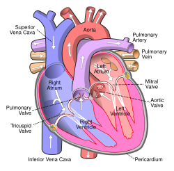 Anterior (frontal) view of the opened heart. White arrows indicate normal blood flow. | |
 Diagram of the alveoli with both cross-section and external view. | |
| Details | |
| Precursor | Truncus arteriosus |
| System | Circulatory system |
| Drains from | Lungs |
| Drains to | Left atrium |
| Artery | Pulmonary artery |
| Identifiers | |
| Latin | venae pulmonales |
| MeSH | D011667 |
| TA98 | A12.3.02.001 |
| TA2 | 4107 |
| FMA | 66643 |
| Anatomical terminology | |

The pulmonary veins are the veins that transfer oxygenated blood from the lungs to the heart. The largest pulmonary veins are the four main pulmonary veins, two from each lung that drain into the left atrium of the heart. The pulmonary veins are part of the pulmonary circulation.
YouTube Encyclopedic
-
1/5Views:107 499866118 32723 981618 052
-
Pulmonary Veins - Location & Function - Human Anatomy | Kenhub
-
Pulmonary Vein Isolation for Atrial Fibrillation
-
Pulmonary Arteries - Location & Function - Human Anatomy | Kenhub
-
Step By Step: Pulmonary Veins Doppler
-
Circulatory System | Pulmonary Circulation
Transcription
Hi, everyone. This is Matt from Kenhub! And in this tutorial, we will discuss the pulmonary veins, their location, and their function. The topic is fairly simple and straightforward with one very important exception. The exception is that, any other time we use the term vein, we’re discussing a vessel that carries deoxygenated blood. But in the case of the pulmonary veins, they are carrying oxygenated blood. There are four pulmonary veins in total, two connected with each lung. The right pulmonary veins, seen here from a posterior view, carry blood from the right lung into the left atrium of the heart where it is distributed to the rest of the circulatory system. They are the right inferior pulmonary vein and the right superior pulmonary vein. The left pulmonary veins carry blood from the left lung into the left atrium of the heart where it continues to flow outward to the rest of the body. The two left pulmonary veins are also named for their location: the left inferior pulmonary vein and the left superior pulmonary vein. Here’s one way to remember. All veins carry blood to the heart, and all arteries carry blood away from the heart, right? Well, it’s the same here except that the pulmonary veins carry blood to the heart from the lungs, so it’s oxygenated. When you hear the term pulmonary vein, think of it as self-describing. Think lung to heart. And that should help jog the memory. This video is more fun than reading a text book, right? If you want more videos, interactive quizzes, articles, and an atlas of human anatomy, click on the “Take me to Kenhub” button. It is time to say goodbye to your old textbooks, and say hello to your new anatomy learning partner, Kenhub. See you there! https://www.kenhub.com
Structure
There are four main pulmonary veins, two from each lung – an inferior and a superior main vein, emerging from each hilum. The main pulmonary veins receive blood from three or four feeding veins in each lung, and drain into the left atrium. The peripheral feeding veins do not follow the bronchial tree. They run between the pulmonary segments from which they drain the blood. [1]
At the root of the lung, the right superior pulmonary vein lies in front of and a little below the pulmonary artery; the inferior is situated at the lowest part of the lung hilum. Behind the pulmonary artery is the bronchus.[2] The right main pulmonary veins (contains oxygenated blood) pass behind the right atrium and superior vena cava; the left in front of the descending thoracic aorta.[citation needed]
Variation
Occasionally the three lobar veins on the right side remain separate, and not infrequently the two left lobar veins end by a common opening into the left atrium. Therefore, the number of pulmonary veins opening into the left atrium can vary between three and five in the healthy population.[citation needed]
The two left lobar veins may be united as a single pulmonary vein in about 25% of enssaam[citation needed]
- the two right veins may be united in about 3%.[2]
Function
The pulmonary veins play an essential role in respiration, by receiving blood that has been oxygenated in the alveoli and returning it to the left atrium.[citation needed]
Clinical significance
As part of the pulmonary circulation they carry oxygenated blood back to the heart, as opposed to the veins of the systemic circulation which carry deoxygenated blood.[citation needed]
On chest X-ray, the diameters of pulmonary veins increases from upper to lower lobes, from 3 mm at the first intercoastal space, to 6 mm just above the diaphragm.[3]
A rare genetic defect of the pulmonary veins can cause them to drain into the pulmonary circulation in whole or in part, this is known as a total anomalous pulmonary venous connection (or drainage), or partial anomalous pulmonary connection, respectively.[citation needed]
Additional images
-
 Computed tomography of a normal lung, with different levels of pulmonary veins.
 -
 Bronchial anatomy
 -
 Transverse section of thorax, showing relations of pulmonary artery.
 -
 Pulmonary vessels, seen in a dorsal view of the heart and lungs.


See also
References
![]() This article incorporates text in the public domain from page 642 of the 20th edition of Gray's Anatomy (1918)
This article incorporates text in the public domain from page 642 of the 20th edition of Gray's Anatomy (1918)
- ^ Drake, Richard L.; Vogl, Wayne; Tibbitts, Adam W.M. Mitchell; illustrations by Richard; Richardson, Paul (2005). Gray's anatomy for students (Pbk. ed.). Philadelphia: Elsevier/Churchill Livingstone. ISBN 978-0-443-06612-2.
- ^ a b Skandalakis, editor in chief John E. (2004). "Chapter 7. Pericardium, Heart, and Great Vessels in the Thorax". Skandalakis' surgical anatomy : the embryologic and anatomic basis of modern surgery. Athens, Greece: PMP. pp. section titled 'Pulmonary veins'. ISBN 9603990744.
{{cite book}}:|first=has generic name (help) - ^ Porres, Diego Varona; Morenza, Óscar Persiva; Pallisa, Esther; Roque, Alberto; Andreu, Jorge; Martínez, Manel (July 2013). "Learning from the Pulmonary Veins". RadioGraphics. 33 (4): 999–1022. doi:10.1148/rg.334125043. ISSN 0271-5333. PMID 23842969.
External links
- Anatomy figure: 19:05-08 at Human Anatomy Online, SUNY Downstate Medical Center
- Illustration at infomat.net