
| Prevertebral space | |
|---|---|
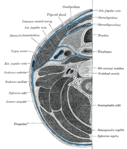 Section of the neck at about the level of the sixth cervical vertebra. Showing the arrangement of the fascia coli. | |
 Sagittal section of nose mouth, pharynx, and larynx. | |
| Anatomical terminology |
The prevertebral space is a space in the neck.
On one side it is bounded by the prevertebral fascia.[1]
On the other side, some sources define it as bounded by the vertebral bodies,[2] and others define it as bounded by the longus colli.[1]
It includes the prevertebral muscles (longus colli and longus capitis), vertebral artery, vertebral vein, scalene muscles, phrenic nerve and part of the brachial plexus.[3]
In trauma, an increased thickness of the prevertebral space is a sign of injury, and can be measured with medical imaging.[4]
YouTube Encyclopedic
-
1/3Views:12 0896 501599 996
-
Neck: Cervical Compartments & Cervical Fascia – Brain & Nervous System | Lecturio
-
The Submandibular Space - Anatomy Tutorial (Mosaiced.org)
-
Neck Anatomy - Organisation of the Neck - Part 1
Transcription
Clinical significance
On plain radiography, prevertebral space should be less than 6 mm at C3 vertebral level in children; while in adults, the space should be less than 6 mm at C2 level and less than 22 mm at C6 level. Causes of enlarged prevertebral space could be edema, hematoma, abscess, tumors, and post surgical changes.[5]
![CT scan with upper limits of the thickness of the prevertebral space at different levels.[4]](/wikipedia/commons/thumb/d/d2/CT_of_prevertebral_space.jpg/151px-CT_of_prevertebral_space.jpg)
References
- ^ a b "eMedicine - Retropharyngeal Abscess : Article by Todd J Berger, MD". Retrieved 2008-02-18.
- ^ "Deep Neck Space Infections: Changing Trends". Archived from the original on March 19, 2007. Retrieved 2008-02-18.
- ^ "Prevertebral space cervical". Medcyclopaedia. GE.[dead link]
- ^ a b Rojas, C.A.; Vermess, D.; Bertozzi, J.C.; Whitlow, J.; Guidi, C.; Martinez, C.R. (2009). "Normal Thickness and Appearance of the Prevertebral Soft Tissues on Multidetector CT". American Journal of Neuroradiology. 30 (1): 136–141. doi:10.3174/ajnr.A1307. ISSN 0195-6108. PMC 7051716.
- ^ Debnam JM, Guha-Thakurta N (December 2012). "Retropharyngeal and prevertebral spaces: anatomic imaging and diagnosis". Otolaryngologic Clinics of North America. 45 (6): 1293–310. doi:10.1016/j.otc.2012.08.004. PMC 3994542. PMID 23153750.
This anatomy article is a stub. You can help Wikipedia by expanding it. |