
| Anterior external arcuate fibers | |
|---|---|
 Diagram showing the course of the arcuate fibers. (Testut.) 1. Medulla oblongata anterior surface. 2. Anterior median fissure. 3. Fourth ventricle. 4. Inferior olivary nucleus, with the accessory olivary nuclei. 5. Gracile nucleus. 6. Cuneate nucleus. 7. Trigeminal. 8. Inferior peduncles, seen from in front. 9. Posterior external arcuate fibers. 10. Anterior external arcuate fibers. 11. Internal arcuate fibers. 12. Peduncle of inferior olivary nucleus. 13. Nucleus arcuatus. 14. Vagus. 15. Hypoglossal. | |
 Section of the medulla oblongata at about the middle of the olive. (Arcuate fibers labeled at center right.) | |
| Details | |
| Identifiers | |
| Latin | fibrae arcuatae externae anteriores |
| NeuroLex ID | birnlex_1628 |
| Anatomical terminology | |
The anterior external arcuate fibers (ventral external arcuate fibers) vary as to their prominence: in some cases they form an almost continuous layer covering the medullary pyramids and olivary body, while in other cases they are barely visible on the surface.
Most of them reach the surface by way of the anterior median fissure, and arch backward over the pyramid.
Reinforced by others which emerge between the pyramid and olive, they pass backward over the olive and lateral district of the medulla oblongata, and enter the inferior peduncle.
As the fibers arch across the pyramid, they enclose a small nucleus which lies in front of and medial to the pyramid.
This is named the arcuate nucleus, and is serially continuous above with the pontine nuclei in the pons; it contains small fusiform (spindle-shaped) cells, around which some of the arcuate fibers end, and from which others arise.
YouTube Encyclopedic
-
1/3Views:4 7052 66724 423
-
Ascending Tract - Dorsal Column/Medial Lemniscus (Neuroanatomy) for USMLE
-
Cuneocerebellar Pathway
-
A brief description of the Spinocerebellar Tracts
Transcription
Additional images
-
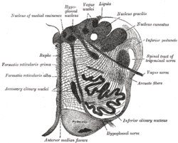
 The reticular formation shown by a transverse section passing through the middle of the olive.

References
![]() This article incorporates text in the public domain from page 782 of the 20th edition of Gray's Anatomy (1918)
This article incorporates text in the public domain from page 782 of the 20th edition of Gray's Anatomy (1918)
External links
This neuroanatomy article is a stub. You can help Wikipedia by expanding it. |