
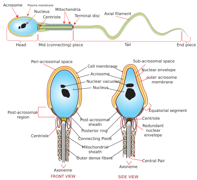
The acrosome is an organelle that develops over the anterior (front) half of the head in the spermatozoa (sperm cells) of humans, and many other animals. It is a cap-like structure derived from the Golgi apparatus. In placental mammals, the acrosome contains degradative enzymes (including hyaluronidase and acrosin).[1] These enzymes break down the outer membrane of the ovum,[2] called the zona pellucida, allowing the haploid nucleus in the sperm cell to join with the haploid nucleus in the ovum. This shedding of the acrosome, or acrosome reaction, can be stimulated in vitro by substances a sperm cell may encounter naturally such as progesterone[3] or follicular fluid, as well as the more commonly used calcium ionophore A23187.[4] This can be done to serve as a positive control when assessing the acrosome reaction of a sperm sample by flow cytometry[5] or fluorescence microscopy. This is usually done after staining with a fluoresceinated lectin such as FITC-PNA, FITC-PSA, FITC-ConA, or fluoresceinated antibody such as FITC-CD46.[6]
In the case of globozoospermia (sperm with round heads), the Golgi apparatus is not transformed into the acrosome, causing male infertility.[7]
YouTube Encyclopedic
-
1/3Views:216 06534 127245 763
-
General Embryology - Detailed Animation On Fertilization
-
Developmental biology part 4 : sea urchin fertilization
-
Egg, sperm, and fertilization | Behavior | MCAT | Khan Academy
Transcription
when ejaculation occurs during intercourse approximately 200 million sperm or spermatozoa are deposited into the vagina they swim through the cervix propelled by with whip-like motions of their tails or Flagela after which muscular contractions of the uterus direct them to the uterine tubes this process usually takes between 30 minutes and two hours only around 200 spermatozoa will reach the secondary oocyte in the uterine tube and of these only one will fertilize it fertilization cannot occur until two processes have taken place capacitation and the acrosomal reaction these can take several hours capacitation is not fully understood but secretions from the uterus wall and uterine tube destabilize the plasma membrane surrounding the head the spermatozoa or acrosome resulting in the membrane becoming more fluid which helps to prepare the spermatozoa for the events a fertilization the spermatozoa become hyperactive their flagella beat more frequently and their heads move laterally the capacitated spermatozoa moved through the corona radiata a dense layer of granulosa cells surrounding the oocyte and come into contact with the zona pellucida the zona pellucida expresses specific receptor proteins called ZP3 which bind to proteins expressed in the heads of the spermatozoa the binding ZP3 triggers the acrosome reaction during which the enzymatic contents of the acrosome are released these enzymes help to digest a path through the zona pellucida allowing the spermatozoa to enter the perivitelline space and reach the plasma membrane of the secondary oocyte with which it fuses to ensure that only one spermatozoa and penetrates the zona pellucida infuses with the oocyte membrane fusion of the spermatozoon and oocyte membranes activates a fast and a slow block to polyspermy during fast blocked polyspermy after fusion the oocyte membrane depolarizes preventing other spermatozoa from fusing with it slow block to polyspermy is also stimulated by this depolarization during slow block to polyspermy a wave of intracellular calcium is released causing small cortical granules beneath the oocyte membrane to release their contents renderings ZP3 inactive and making the zona pellucida impermeable upon the spermatozoon entering the oocyte undergoes meiosis II and further develops into the female pro nucleus during this time the sperm develops into the male pro nucleus and the two Pro nuclei fuse to form a single diploid nucleus or zygote
See also
References
- ^ "acrosome definition - Dictionary - MSN Encarta". Archived from the original on 2009-02-14. Retrieved 2007-08-15.
- ^ Larson, Jennine L.; Miller, David J. (1999). "Simple histochemical stain for acrosomes on sperm from several species". Molecular Reproduction and Development. 52 (4): 445–449. doi:10.1002/(SICI)1098-2795(199904)52:4<445::AID-MRD14>3.0.CO;2-6. ISSN 1098-2795. PMID 10092125. S2CID 24542696.
- ^ Lishko, Polina V.; Botchkina, Inna L.; Kirichok, Yuriy (March 2011). "Progesterone activates the principal Ca 2+ channel of human sperm". Nature. 471 (7338): 387–391. Bibcode:2011Natur.471..387L. doi:10.1038/nature09767. ISSN 1476-4687. PMID 21412339. S2CID 4340309.
- ^ Jamil, K.; White, I. G. (December 1981). "Induction of acrosomal reaction in sperm with ionophore A23187 and calcium". Archives of Andrology. 7 (4): 283–292. doi:10.3109/01485018108999319. ISSN 0148-5016. PMID 6797354.
- ^ Miyazaki R, Fukuda M, Takeuchi H, Itoh S, Takada M (1990). "Flow cytometry to evaluate acrosome-reacted sperm". Arch. Androl. 25 (3): 243–51. doi:10.3109/01485019008987613. PMID 2285347.
- ^ Carver-Ward JA, Moran-Verbeek IM, Hollanders JM (February 1997). "Comparative flow cytometric analysis of the human sperm acrosome reaction using CD46 antibody and lectins". J. Assist. Reprod. Genet. 14 (2): 111–9. doi:10.1007/bf02765780. PMC 3454831. PMID 9048242.
- ^ Hermann Behre; Eberhard Nieschlag (2000). Andrology : Male Reproductive Health and Dysfunction. Berlin: Springer. p. 155. ISBN 3-540-67224-9.
This article related to the genitourinary system is a stub. You can help Wikipedia by expanding it. |