
| Vitelline veins | |
|---|---|
 | |
| Details | |
| Carnegie stage | 9 |
| Days | 28 |
| Identifiers | |
| Latin | vena vitellina |
| FMA | 70305 |
| Anatomical terminology | |
The vitelline veins are veins that drain blood from the yolk sac[1] and the gut tube[2] during gestation.
YouTube Encyclopedic
-
1/5Views:7555 21425 338184 777109 258
-
Embryology of Vitelline veins (Portal vein development ) | Med Bees
-
Development of Veins Associated With Heart [Part 1]
-
Development of veins - Dr. Ahmed Farid
-
Embryology | Development of Vascular System
-
Medical Embryology - Development of Cardinal Veins and the Large Veins
Transcription
Path
They run upward at first in front, and subsequently on either side of the intestinal canal. They unite on the ventral aspect of the canal.
Beyond this, they are connected to one another by two anastomotic branches, one on the dorsal, and the other on the ventral aspect of the duodenal portion of the intestine. This is encircled by two venous rings; into the middle or dorsal anastomosis the superior mesenteric vein opens.
The portions of the veins above the upper ring become interrupted by the developing liver and broken up by it into a plexus of small capillary-like vessels termed sinusoids.
Derivatives

The vitelline veins give rise to:[4]
- Hepatic veins
- Inferior portion of Inferior vena cava
- Portal vein
- Superior mesenteric vein
- Inferior mesenteric vein
The branches conveying the blood to the plexus are named the venae advehentes, and become the branches of the portal vein. The vessels draining the plexus into the sinus venosus are termed the venae revehentes, and form the future hepatic veins.[3] Ultimately the left vena revehens no longer communicates directly with the sinus venosus, but opens into the right vena revehens. The persistent part of the upper venous ring, above the opening of the superior mesenteric vein, forms the trunk of the portal vein.
Function
The vitelline veins drain the yolk sac during early embryonic development.[1][5] They also drain the gut tube in embryos once this has formed from the yolk sac.[2][6]
Additional images
-
 Chick embryo of thirty-three hours’ incubation, viewed from the dorsal aspect. X 30.
 -
 Model of human embryo 1.3 mm. long.
 -
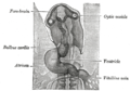 Head of chick embryo of about thirty-eight hours’ incubation, viewed from the ventral surface. X 26.
 -
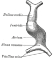 Diagram to illustrate the simple tubular condition of the heart.
 -
 Liver with the septum transversum. Human embryo 3 mm. long.


References
- ^ a b Carlson, Bruce M. (2014-01-01), "Development of the Vascular System", Reference Module in Biomedical Sciences, Elsevier, doi:10.1016/b978-0-12-801238-3.05459-3, ISBN 978-0-12-801238-3, retrieved 2020-11-14
- ^ a b Snarr, Brian S.; McQuinn, Tim C.; Wessels, Andy (2017-01-01), Polin, Richard A.; Abman, Steven H.; Rowitch, David H.; Benitz, William E. (eds.), "50 - Cardiovascular Development", Fetal and Neonatal Physiology (Fifth Edition), Elsevier, pp. 515–522.e2, doi:10.1016/b978-0-323-35214-7.00050-0, ISBN 978-0-323-35214-7, retrieved 2020-11-14
- ^ a b Nagy, Rodica Daniela; Ruican, Dan; Zorilă, George-Lucian; Istrate-Ofiţeru, Anca-Maria; Badiu, Anne Marie; Iliescu, Dominic Gabriel (February 2022). "Feasibility of Fetal Portal Venous System Ultrasound Assessment at the FT Anomaly Scan". Diagnostics. 12 (2): 361. doi:10.3390/diagnostics12020361. PMC 8871164. PMID 35204452.
- ^ "Vitelline veins: Derivatives". LifeHugger. Archived from the original on 2012-03-05. Retrieved 2009-12-11.
- ^ Maynard, Robert Lewis; Downes, Noel (2019-01-01), Maynard, Robert Lewis; Downes, Noel (eds.), "Chapter 14 - Liver", Anatomy and Histology of the Laboratory Rat in Toxicology and Biomedical Research, Academic Press, pp. 159–168, doi:10.1016/b978-0-12-811837-5.00014-9, ISBN 978-0-12-811837-5, retrieved 2020-11-14
- ^ Mitchell, Barry; Sharma, Ram (2009-01-01), Mitchell, Barry; Sharma, Ram (eds.), "Chapter 6 - The cardiovascular system", Embryology (Second Edition), Churchill Livingstone, pp. 31–40, doi:10.1016/b978-0-7020-3225-7.50009-9, ISBN 978-0-7020-3225-7, retrieved 2020-11-14
External links
- Embryology at Temple Heart98/heart97a/sld020
- cardev-009—Embryo Images at University of North Carolina
- cardev-016—Embryo Images at University of North Carolina