
| Foramen ovale (heart) | |
|---|---|
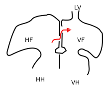 Sketch showing foramen ovale in a fetal heart. Red arrow shows blood from the inferior cava traveling to the right atrium and then to the left atrium. HF: right atrium, VF: left atrium. HH and VH: right and left ventricle. The heart still has a common pulmonary vein (LV), instead of four. | |
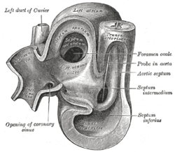 Heart of human embryo of about thirty-five days, opened on left side. | |
| Details | |
| Precursor | Septum secundum |
| System | Cardiovascular system |
| Identifiers | |
| MeSH | D054085 |
| TA98 | A12.1.01.007 |
| TA2 | 3967 |
| FMA | 86043 |
| Anatomical terminology | |
In the fetal heart, the foramen ovale (/fəˈreɪmən oʊˈvæli, -mɛn-, -ˈvɑː-, -ˈveɪ-/[1][2][3]), also foramen Botalli or the ostium secundum of Born, allows blood to enter the left atrium from the right atrium. It is one of two fetal cardiac shunts, the other being the ductus arteriosus (which allows blood that still escapes to the right ventricle to bypass the pulmonary circulation). Another similar adaptation in the fetus is the ductus venosus. In most individuals, the foramen ovale closes at birth. It later forms the fossa ovalis.
YouTube Encyclopedic
-
1/3Views:269 82111 64225 258
-
Foramen ovale and ductus arteriosus | Circulatory system physiology | NCLEX-RN | Khan Academy
-
What's Patent Foramen Ovale (PFO): Pediatrics
-
Patent Foramen Ovale Closure
Transcription
What you are looking at is the fetal heart Looks a lot like a human heart but a couple of interesting differences that we are gonna go over Uh the first thing I want you to know is that its mostly got what I've drawn is a kind of a purplish blood In the adult heart, we know that there is a real clear distinction between oxygenated blood and deoxygenated blood In the fetal heart, it's all very very similar Now, let's start by kind of orienting ourselves, this vessel at the top is different It has got blue blood rather than a purplish blood and the reason for that is that this blood is actually coming back from the body and the body has used up as much oxygen as it can so this is the Superior Vena Cava, dragging blood back from the arms, and specifically the head region. And you've also got, from the bottom, blood coming into the heart via the Inferior Vena Cava. Now this is also coming from the body, but I've drawn it more pink. So why would I do that? Well, it's because, if you remember, there's also in addition to just bringing blood from the body There is also blood coming from the Umbilical vein. And I don't want you to forget that because the umbilical vein is actually bringing really really oxygen rich blood from the placenta and its mixing with the inferior vena cava. So it's not bright red, but it has got this kind of pinkish tone to it and this is really the only major source of oxygen that fetus is getting from this umbilical vein So this is actually very very important and thats why when it mixes with the blue blood from the superior vena cava in the right atrium we get this purplish stuff... and of course, let me just quickly label the rest of the chambers you've got the right ventricle the left atrium and the left ventricle and these are the four chambers, right? let me also name the major arteries and veins this, of course, is the aorta at the top I've also got the pulmonary artery here Pulmonary artery and we've got the Pulmonary veins and I am just going to label this side, right here "Pulmonary Veins". But you can see there are two (2) on the other side as well. So, this is what the fetal heart looks like and now actually think about what's going on in the fetal heart and how the blood is flowing through So to do that, let me actually start out by drawing some lungs This is actually going to help inform the path of blood So these are the lungs Let's say this is the right lung... and of course, there is one on the left as well let me just draw it in just so we don't forget that it exists But I'm going to use the right lung for this example This is our left lung; I even drew the little cardiac notch And on the right lung, blood is coming in from, the pulmonary artery So blood is coming in this way from the pulmonary artery It's going to go into little vessels little arterioles I'll draw them in the same purplish colour It's going to get into the little arterioles, and then get into the little capillaries. Even tinier little blood vessels and those capillaries are gonna go and meet up with an alveolar sac and these sacs, in adults, are full of air. But in the fetus, there is actually nothing but fluid inside So it's actually just full of amniotic fluid It's just fluid filled so if you are thinking about it do you expect the oxygen level to be high, or low? Well, if it's full of fluid, amniotic fluid, it's going to be pretty low There's not much oxygen in there In fact, blood isn't even going to the lungs to get oxygen because we said that the main source of oxygen for the fetus is going to be from the umbilical vein This is where the vast majority of the oxygen is coming from So, what ends up happening is that because there is such low oxygen in the alveolar sacs they have this process, this ability to actually cause the arterioles to constrict these arterioles have some smooth muscles on them and they actually the alveolar sacs because of low oxygen they make this constrict so literally kind of clamps down and it looks a little tighter, something like this... a skinnier blood vessel and when it constricts when you have a smaller, you know, radius on that blood vessel, what does that mean exactly? Well that means that the amount of resistance went up here and, of course, if it happens once, it's not a big deal but if it happens in millions and millions of arterioles all throughout the lungs then what we are really talking about is that the pulmonary artery, both of them on both the sides are going to face really high resistance. and this process of increasing the resistance when the amount of oxygen is low remember we actually named this process this process is called "Hypoxic.." (that just means low oxygen) Pulmonary (referring to the lungs) Vaso (meaning blood vessel) constriction (so making the blood vessels tight) So, this is the process we are talking about 'Hypoxic Pulmonary Vasoconstriction and it happens in adults, but it also happens in the fetus it is actually very important in the fetus because the lungs, both the left and right are full of fluid it allows the blood vessels to constrict It causes the blood vessels to constrict and it really raises the amount of resistance that the pulmonary arteries are facing now, if they are facing a lot of resistance think about what that means That means that if the heart wants to pump blood to the lungs, it's going to have to raise the pressure So the pressure goes up in the pulmonary artery and if the pressure is high there, that means the pressure is going to be high in the right ventricle and if the pressure is high in the right ventricle, blood has to get in there somehow so the pressure starts going up in the right atrium so pressures start going up everywhere and, so really, what the heart faces, is a choice. They can either continue to just try to push blood into the lungs, even though there is a lot of resistance, or, it can try to find a short-cut really to by pass the lungs altogether. And that idea of short-cuts is really what we are talking about in the fetal heart In fact, there are two (2). there are two short cuts to get blood from this side the right side, either the right atrium, or the right ventricle or the pulmonary artery over to the left side and when I say left side, I really mean at the end of the day the aorta or the left atrium or the left ventricle as well But really, at the end of the day, you want to get blood into the aorta and you want to think of a clever way of doing it and being able to bypass the lungs That's the challenge. So, how does the fetal heart meet that challenge? how does it bypass the lungs? two major ways So, let me draw them both out and I am going to start by drawing a blow-up of this section right here Lets say I blow that up and I am going to try to sketch it out here for you let's see if I can make it neat this is the same box (just want to make sure we are not confused by the way I am drawing it) this is just a blow-up of that section and if you look closely what you would see is that there is a wall the same wall that I drew but there is actually not just one wall, there is two walls stuck together thats actually the first point I want to make is that there is not just one, but two walls there and this one is called septum primum 'Septum' refers to 'a wall' and 'primum' is the latin word for 'first' and so septum primum is this wall over here on this side and Septum Secundum, is the other wall so you've got two walls next to each other and they look almost like one wall but there is actually two and that's the 'Septum Secundum' and just to make sure we are oriented to the right and left atrium on this side is the right atrium and on this side is the left atrium and remember, the overall goal is to somehow bypass the lungs and what I mean is get blood from the here, somehow over to the other side and what happens is that when you look closely at this Septum Secundum there is a little, tiny hole there so little bit like a imagine a piece of swiss cheese and if I was looking at this wall as if it's a piece of swiss cheese the hole is actually in the wall so there is a little hole there and if I stuck my finger in there let's stay I stuck my finger right here I would actually be able to touch, from the right atrium I can actually touch the Septum Primum because of the fact that there is a hole in the wall and so this hole is called the 'Foramen Ovale' . Let me just label that for you So the Foramen Ovale is this hole You can now call it by its full name and this Foramen Ovale is right there It turns out that the Septum primum is also like a piece of swiss cheese both of them are like little pieces of swiss cheese and it's got a little break in its wall as well now, think about it, what will happen if the pressure is really high in the right atrium pressure is really high on this side and ofcourse, pressure is the force pushing on the area the surface area of the chamber so blood is pushing in all directions and when it pushes here, right in the Foramen Ovale, what is going to happen? Well, right at that spot if there is pressure, then this Septum Primum does an interesting thing , it becomes a little bit like a flap Or a valve, and it kind of falls away It falls away like that and so Bingo! you've got access Right atrium's blood is going to flow right into the left atrium because the Septum Primum became a little flap and fell away so let me re-sketch this in the middle drawing and make it the way it should be drawn so instead of drawing it like that you've got, literally, a little flap here this is the Septum Primum flap of tissue and you've got your Septum Secundum and I'm going to draw blood now going through so you've got blood flowing through blood is now going to go through from here into the left atrium and remember, this little hole ( I cant really draw a hole very easily , but you can imagine there is a hole there) in the wall of the Septum Secundum, where the blood is going through that hole is called the Foramen Ovale and so this is kind of when I said there are a couple of tricks This is trick number 1. trick # 1 is getting blood from the right atrium directly over to the left atrium because you know that once it gets over there now it has literally just bypassed the lungs but that's only one of two tricks Not all of the blood in the right atrium goes through the foramen ovale not all of it Some blood actually passes through the normal way it goes to the tricuspid valve into the right ventricle and if blood is going to the right ventricle that is a good thing Why? because we want to make sure our right ventricle is pumping. We want to make sure that it is squeezing getting practice and that those muscles are getting stronger Now, the right ventricle is going to do its job its going to pump blood into the Pulmonary Arteries the left and right pulmonary arteries and that blood is facing as I said before a lot of resistance So, there is another little trick turns out, there is another place for the blood to go you are looking at this picture, thinking, "Well, I don't see any other place for blood to go" "There's only the left pulmonary artery and the right pulmonary artery" but there is another place turns out that the fetal heart actually has a little vessel here I am going to do the vessel in another colour and this vessel allows blood to go through so you can actually get blood to pass through this vessel like so so, this is actually really cool, right because you can now see how blood can go directly from the pulmonary artery into the aorta and go down this is our trick # 2 so this little vessel, this little guy Im going to loop it name it, this is our Ductus Arteriosis now, there is another one called the Ductus venosus , a diifferent name but this is trick number 2 one trick was to go from the right atrium to the left atrium and another trick was to go from the pulmonary arteries to the aorta so these are the two major tricks and you basically can see now that blood is going to bypass the lungs using either trick. now does that mean that no blood goes to the lungs? No. A little bit of blood does go to the lungs and in fact about 10% , or so, continues to the lungs but 90% actually goes through one of these two pathways. either through the Ductus or through the Foramen Ovale. So these are the interesting differences between the fetal heart and the adult heart.
Development
The foramen ovale (from Latin 'oval hole') forms in the late fourth week of gestation, as a small passageway between the septum secundum and the ostium secundum. Initially the atria are separated from one another by the septum primum except for a small opening below the septum, the ostium primum. As the septum primum grows, the ostium primum narrows and eventually closes. Before it does so, bloodflow from the inferior vena cava wears down a portion of the septum primum, forming the ostium secundum. Some embryologists postulate that the ostium secundum may be formed through programmed cell death.[4]
The ostium secundum provides communication between the atria after the ostium primum closes completely. Subsequently, a second wall of tissue, the septum secundum, grows over the ostium secundum in the right atrium. Blood then passes from the right to left atrium only by way of a small passageway in the septum secundum and then through the ostium secundum. This passageway is called the foramen ovale.[citation needed]
Closure
The foramen ovale often closes at birth. At birth, when the lungs become functional, the pulmonary vascular pressure decreases and the left atrial pressure exceeds that of the right. This forces the septum primum against the septum secundum, functionally closing the foramen ovale. In time the septa eventually fuse, leaving a remnant of the foramen ovale, the fossa ovalis.
Function
A fetus receives oxygen not from its lungs, but from the mother's oxygen-rich blood via the placenta. Oxygenated blood from the placenta travels through the umbilical cord to the right atrium of the fetal heart. As the fetal lungs are non-functional at this time, the blood bypasses them through two cardiac shunts. The first is the foramen ovale (the valve present between them called eustachian valve) which shunts blood from the right atrium to the left atrium. The second is the ductus arteriosus which shunts blood from the pulmonary artery (which, after birth, carries blood from the right side of the heart to the lungs) to the descending aorta.[citation needed]
Clinical significance
In about 25% of adults the foramen ovale does not close completely, but remains as a small patent foramen ovale ("PFO").[5] In most of these individuals, the PFO causes no problems and remains undetected throughout life.
PFO has long been studied because of its role in paradoxical embolism (an embolism that travels from the venous side to the arterial side). This may lead to a stroke or transient ischemic attack. Transesophageal echocardiography is considered the most accurate investigation to demonstrate a patent foramen ovale. A patent foramen ovale may also be an incidental finding.
See also
References
- ^ "foramen". Merriam-Webster.com Dictionary. Retrieved 2016-01-22. "ovale". Merriam-Webster.com Dictionary. Retrieved 2016-01-22.
- ^ "foramen". Lexico UK English Dictionary. Oxford University Press. Archived from the original on 2020-09-30.
- ^ "foramen". Dictionary.com Unabridged (Online). n.d. Retrieved 2016-01-22.
- ^ Sadler, Thomas W. (2004). Langman's Essential Medical Embryology. Lippincott Williams & Wilkins. ISBN 0-7817-5571-9.
- ^ Homma, S. (2005). "Patent Foramen Ovale and Stroke". Circulation. 112 (7): 1063–1072. doi:10.1161/CIRCULATIONAHA.104.524371. ISSN 0009-7322. PMC 3723385. PMID 16103257.
- Carlson, Bruce (2004). Human Embryology And Developmental Biology (3rd ed.). Elsevier Mosby. ISBN 0-323-03649-X.
- "Congenital Heart Disorders". Cleveland Clinic.