
| Pons | |
|---|---|
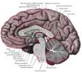
 Pons in the brainstem | |
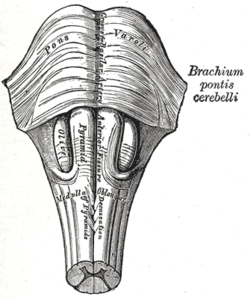 Anteroinferior view of the medulla oblongata and pons | |
| Details | |
| Part of | Brain stem |
| Artery | Pontine arteries |
| Vein | Transverse and lateral pontine veins |
| Identifiers | |
| MeSH | D011149 |
| NeuroNames | 547 |
| NeuroLex ID | birnlex_733 |
| TA98 | A14.1.03.010 |
| TA2 | 5921 |
| FMA | 67943 |
| Anatomical terms of neuroanatomy | |
The pons (pl.: pontes; from Latin pons, "bridge", from Proto-Indo-European *pónteh₁s, “path, road”, from *pent-, “path”. Cognate with Sanskrit पन्था, pánthā-) is part of the brainstem that in humans and other mammals, lies inferior to the midbrain, superior to the medulla oblongata and anterior to the cerebellum.
The pons is also called the pons Varolii ("bridge of Varolius"), after the Italian anatomist and surgeon Costanzo Varolio (1543–75).[1] This region of the brainstem includes neural pathways and tracts that conduct signals from the brain down to the cerebellum and medulla, and tracts that carry the sensory signals up into the thalamus.[2]
YouTube Encyclopedic
-
1/5Views:202 15994 192224 38470 815121 886
-
Neurology | Pons Anatomy & Function
-
Pons - External and Internal (White & Grey matter) + QUIZ | Anatomy
-
Pons - Structure & Function - Neuroanatomy
-
Pons Lesions
-
Pons Anatomy (1/3) | External Features
Transcription
Structure
The pons in humans measures about 2.5 centimetres (0.98 in) in length.[2] It is the part of the brainstem situated between the midbrain and the medulla oblongata,[3] and in front of the cerebellum.[citation needed] The horizontal medullopontine sulcus demarcates the boundary between the pons and medulla oblongata on the ventral aspect of the brainstem, and the roots of cranial nerves VI/VII/VIII emerge from the brainstem along this groove.[4] The junction of pons, medulla oblongata, and cerebellum forms an angle - the cerebellopontine angle.[5] The superior pontine sulcus separates the pons from the midbrain.[6] Posteriorly, the pons curves on either side into a middle cerebellar peduncle.[3]
The pons can be broadly divided into two parts: the basilar part of the pons (ventral pons),[7] and the pontine tegmentum (dorsal pons).[8]
The ventral aspect of the pons faces the clivus, with the pontine cistern intervening between the two structures. The ventral surface of the pons features a midline basilar sulcus along which the basilar artery may or may not course. There is a bulge to either side of the basilar sulcus, created by the pontine nuclei that are interweaved amid the descending fibres within the substance of the pons. The superior cerebellar artery winds around the upper margin of the pons.[3]
Vasculature
Most of the pons is supplied by the pontine arteries, which arise from the basilar artery. A smaller portion of the pons is supplied by the anterior and posterior inferior cerebellar arteries.
Development
During embryonic development, the metencephalon develops from the rhombencephalon and gives rise to two structures: the pons and the cerebellum.[2] The alar plate produces sensory neuroblasts, which will give rise to the solitary nucleus and its special visceral afferent (SVA) column; the cochlear and vestibular nuclei, which form the special somatic afferent (SSA) fibers of the vestibulocochlear nerve, the spinal and principal trigeminal nerve nuclei, which form the general somatic afferent column (GSA) of the trigeminal nerve, and the pontine nuclei which relays to the cerebellum.
Basal plate neuroblasts give rise to the abducens nucleus, which forms the general somatic efferent fibers (GSE); the facial and motor trigeminal nuclei, which form the special visceral efferent (SVE) column, and the superior salivatory nucleus, which forms the general visceral efferent fibers (GVE) of the facial nerve.
Nuclei

A number of cranial nerve nuclei are present in the pons:
- mid-pons: the principal sensory nucleus of the trigeminal nerve (V)
- mid-pons: the motor nucleus for the trigeminal nerve (V)
- lower down in the pons: abducens nucleus (VI)
- lower down in the pons: facial nerve nucleus (VII)
- lower down in the pons: vestibulocochlear nuclei (vestibular nuclei and cochlear nuclei) (VIII)
Function
Functions of these four cranial nerves (V-VIII) include regulation of respiration, control of involuntary actions, sensory roles in hearing, equilibrium, and taste, and in facial sensations such as touch and pain, as well as motor roles in eye movement, facial expressions, chewing, swallowing, and the secretion of saliva and tears.[2]
The pons contains nuclei that relay signals from the forebrain to the cerebellum, along with nuclei that deal primarily with sleep, respiration, swallowing, bladder control, hearing, equilibrium, taste, eye movement, facial expressions, facial sensation, and posture.[2]
Within the pons is the pneumotaxic center consisting of the subparabrachial and the medial parabrachial nuclei. This center regulates the change from inhalation to exhalation.[2]
The pons is implicated in sleep paralysis, and may also play a role in generating dreams.[9]
Clinical significance
- Central pontine myelinolysis is a demyelinating disease that causes difficulty with sense of balance, walking, sense of touch, swallowing and speaking. In a clinical setting, it is often associated with transplant or rapid correction of blood sodium. Undiagnosed, it can lead to death or locked-in syndrome.
Other animals
Evolution
The pons first evolved as an offshoot of the medullary reticular formation.[10] Since lampreys possess a pons, it has been argued that it must have evolved as a region distinct from the medulla by the time the first agnathans appeared, 525 million years ago.[11]
Additional images
-
 Location and topography of pons (animation)
 -
 Axial section of the pons, at its upper part
 -
 Hind- and mid-brains; posterolateral view
 -
 Median sagittal section of brain
 -
 Nuclei of the pons and brainstem
 -
 Cerebrum. Deep dissection. Inferior dissection.


References
- ^ Gray, Henry (1862). Anatomy, descriptive and surgical. Blanchard and Lea. pp. 514–. Retrieved 10 November 2010.
- ^ a b c d e f Saladin, Kenneth S. (2007). Anatomy & physiology the unity of form and function. Dubuque, Iowa: McGraw-Hill.
- ^ a b c Sinnatamby, Chummy S. (2011). Last's Anatomy (12th ed.). p. 478. ISBN 978-0-7295-3752-0.
- ^ "sulcus bulbopontis". TheFreeDictionary.com. Retrieved 8 June 2023.
- ^ "cerebellopontile angle". TheFreeDictionary.com. Retrieved 8 June 2023.
- ^ Carpenter, M (1985). Core text of neuroanatomy (3rd ed.). Williams & Wilkins. p. 42. ISBN 0683014552.
- ^ "pars basilaris pontis". TheFreeDictionary.com. Retrieved 8 June 2023.
- ^ "tegmentum pontis". TheFreeDictionary.com. Retrieved 8 June 2023.
- ^ Koch, Christof. "Dream States: A Peek into Consciousness". Scientific American. Scientific American. Retrieved 17 September 2020.
- ^ Pritchard and Alloway Medical Neuroscience
- ^ Butler and Hodos Comparative vertebrate neuroanatomy: evolution and adaptation
- Pritchard, TE & Alloway, D (1999). Medical neuroscience. Hayes Barton Press. ISBN 978-1-59377-200-0.
- Butler, AB & Hodos, W (2005). Comparative vertebrate neuroanatomy: evolution and adaptation. Wiley-Blackwell. ISBN 978-0-471-21005-4.