
| Respiratory center | |
|---|---|
 Respiratory groups in the respiratory center and their influence | |
| Identifiers | |
| MeSH | D012125 |
| Anatomical terminology | |
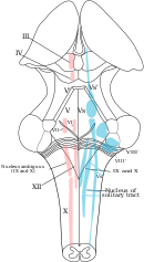
The respiratory center is located in the medulla oblongata and pons, in the brainstem. The respiratory center is made up of three major respiratory groups of neurons, two in the medulla and one in the pons. In the medulla they are the dorsal respiratory group, and the ventral respiratory group. In the pons, the pontine respiratory group includes two areas known as the pneumotaxic center and the apneustic center.
The respiratory center is responsible for generating and maintaining the rhythm of respiration, and also of adjusting this in homeostatic response to physiological changes. The respiratory center receives input from chemoreceptors, mechanoreceptors, the cerebral cortex, and the hypothalamus in order to regulate the rate and depth of breathing. Input is stimulated by altered levels of oxygen, carbon dioxide, and blood pH, by hormonal changes relating to stress and anxiety from the hypothalamus, and also by signals from the cerebral cortex to give a conscious control of respiration.
Injury to respiratory groups can cause various breathing disorders that may require mechanical ventilation, and is usually associated with a poor prognosis.
YouTube Encyclopedic
-
1/5Views:403 947786 892535 109255 011124 796
-
The respiratory center | Respiratory system physiology | NCLEX-RN | Khan Academy
-
Control Of Respiration (regulation of breathing)
-
Respiratory | Regulation of Breathing: Respiratory Centers: Part 1
-
Control of Ventilation, Animation
-
Neural Control of Breathing | Respiratory System
Transcription
So in this video what I wanted to focus on is one particular area of the brain-- actually, two areas I'm going to sketch out, not just one. I'm going to show you how I think that these two areas can be kind of united in a way. So, these two areas, where I'm sketching out these little green circles, are going to be responsible for breathing-- how fast you breathe, how deep you breathe. And there are lots of little neurons in these two areas. And these neurons are going to be communicating. Let's say this neuron sends a little axon on down here, maybe this guy sends an axon up here. They're going to be communicating information among themselves and between themselves to kind of make sure they're working in a coordinated way so that the breathing that you do is the way that it should be, you know, how fast it should be, given a particular situation. So the way I think of it is kind of uniting these two areas. In fact, sometimes it's subdivided even further. I just put it all together and say this part of the brain that I'm just sketching out in green, this area then, is our respiratory center. This is going to be responsible for all of the important activities of breathing. So let me just write that out here. Respiratory center. So our respiratory center is going to gather information from different places. And then it's going to have to make a decision and execute based on all the information it receives. So one key piece of information is going to come from cells right here, neighborhood cells. And these cells are called the central chemoreceptors. The reason I'm calling them central is because they're also part of the brain, right? They're right in the same neighborhood, and so these central chemoreceptors don't have to go too far to communicate their information. And specifically they're going to gather information on things like carbon dioxide levels and pH levels. One thing they don't do is oxygen levels. So that's these guys right here. So if you have central chemoreceptors you also probably can expect that there would be some peripheral chemoreceptors, and these ones are also very important. And they exist outside of the brain, so they're going to be actually sending their information along through neurons that are going to extend all the way into the brain. So for example, you might have two key groups. One is called the aortic body and the other is called the carotid body. Aortic body and the carotid body. They're coming from different locations and are actually going to use different nerves to get into the brain. So the carotid body, for example, is going to extend out this way through a neuron. And that's going to be through a nerve called the-- right here-- called the glossopharyngeal nerve. This is cranial nerve number nine, also called the glossopharyngeal nerve. So this is one of the key peripheral chemoreceptors. You've also got some nerves or neurons projecting from the aortic body going through the vagus nerve. So this is our vagus nerve, or cranial nerve number 10. It goes by two different names. These peripheral chemoreceptors are going to detect things like oxygen-- in fact, that's probably one of the most important things they detect-- as well as carbon dioxide and pH. So that's information coming to the respiratory center from our peripheral and central chemoreceptors then is mostly about chemicals. In addition, there's another whole group of receptors called mechanoreceptors. And these ones are actually going to be sending information about pressure. Now you may be thinking, well, wait a second. I thought baroreceptors told us about pressure. And it turns out, baroreceptors are one type of mechanoreceptor that's found inside of the blood vessels. So there are many other types and many other locations. And so the bigger, more general term would be mechanoreceptor. You can find them in places like the nose, you can find them in the lungs, in the GI tract, so lots of different locations for these mechanoreceptors. And they're all sending their own projection over to the respirator center. And in fact, the lungs and the GI tract are going to hitch a ride in this vagus nerve and the nose mechanoreceptors they're going to travel through another nerve that's called the trigeminal nerve or cranial nerve number five. So these are the routes that these receptors are going to take to get to that respiratory center. But how do these work exactly, these mechanoreceptors? Let's take an example. Let's say you're walking and you inhale some pollen. Well, that's going to trigger one of these mechanoreceptors in your nose and it's going to want to relay that information over to your respiratory center so you're going to get a little nerve impulse through that cranial nerve number five. Similarly in your lungs, let's say you actually inhale some cigarette smoke. And let's say the lungs don't like that. And then the mechanoreceptor feels that little particle. It's going to trigger cranial nerve number 10, the vagus nerve. Similarly, you have these stretch receptors that are in the lungs. And these are actually kind of interesting because what they're doing is they're saying, hey, you know these lungs are starting to get really, really full, really distended, and so they want to let the respiratory center know that maybe it's time to exhale. And similarly, in the GI tract you can imagine, let's say a baby is taking milk and the stomach is getting really distended, you might also imagine that that information would go back to the respiratory center as well, in this case through cranial nerve number 10. So we have information about pressure or stretch, we also have information about chemicals coming in. But what about information on things like, I don't know, things like anxiety for instance, or fear. Let's say someone is having these emotions. Their breathing pattern may change. Maybe they're in pain. So these kinds of things are actually coming from the hypothalamus. So this is another region of the brain that's sending information down to the respiratory center and helping to affect how we breathe. And finally, this is probably the largest part of our picture, this is our cerebrum. And the cerebrum is responsible for all the voluntary stuff that we do, things like singing, where you've got to control your breath. Or maybe you're playing a musical instrument, or maybe you're yelling or screaming. Let's put yelling down here. Anything like that, you're going to want to control your breath. And so that's all voluntary control. So this is our voluntary control. And it's good that actually we have this mechanism so we can-- if we want to-- we can change our breathing pattern. But it's also great that our respiratory center can work on its own. Can you imagine if you had to always think about taking a breath? You couldn't do anything else, right? You couldn't sleep, you couldn't eat. You would always just be thinking about taking a breath so that you wouldn't miss the next breath and subsequently run out of air. So this is all the information coming into our respiratory center. Let me just scoot this over and actually show you now what our brain can do with that information to actually make sure that we're breathing comfortably. This is our spinal column. And I'm actually just going to label out the motor nerves and some of the muscle groups. So we've got motor nerves and muscle groups. And there are four key muscle groups that are going to be controlled by our respiratory center. And we're going to go through them one by one. So the first one, and the one that people usually always talk about or think about, is this one right here. This is going to be C3, C4, and C5. So C3 through C5, and the muscle is the diaphragm. This is the giant muscle that kind of sits right below our lungs and when it contracts you take in a nice deep breath. But it doesn't work alone. We've got other muscles involved as well. So I'm actually going to sketch out what these other muscle groups are. The first one is T1 through T11. All these levels are going to send off a little nerve. And each nerve will go through a different intercostal muscle. So intercostal muscles are-- these are the muscles that kind of go between the ribs-- these are going to help expand or pull out your ribs, right? So these are important for breathing as well. A little bit lower then, you also have these abdominal muscles. Abdominal muscles here are going to be T6 through L1. These are the levels where the little nerve fibers come out and are going to help innervate or help these muscles, abdominal muscles, contract. So this is a third group of muscles and they're controlled by these spinal levels. And the final group would be this group up here. So this is actually C1 through C3. And these would be the accessory muscles. Accessory muscles are the ones-- usually I think of them as the ones around your neck area. And they're going to also kind of help pull out the rib cage and expand the lungs. So there you have it. You have information coming in, that's the stuff that we started talking about, from all the different locations, around chemicals, information about pressure, and your emotional status, and what you're thinking about doing involuntarily. All that information is going to come in and then the respiratory center has to decide how to kind of balance all that information. And on the way out it's going to be able to execute by controlling all these different muscle groups and sending information down the motor nerves that we just listed to these four big groups of muscles.
Respiratory groups
The respiratory center is divided into three major groups, two in the medulla and one in the pons. The two groups in the medulla are the dorsal respiratory group and the ventral respiratory group. In the pons, the pontine respiratory group is made up of two areas – the pneumotaxic center and the apneustic center. The dorsal and ventral medullary groups control the basic rhythm of respiration.[1][2] The groups are paired with one on each side of the brainstem.[3]
Dorsal respiratory group
The dorsal respiratory group (DRG) has the most fundamental role in the control of respiration, initiating inspiration (inhalation). The DRG is a collection of neurons forming an elongated mass that extends most of the length of the dorsal medulla. They are near to the central canal of the spinal cord, and just behind the ventral group. They set and maintain the rate of respiration.[4][5]
Most of the neurons are located in the nucleus of the solitary tract. Other important neurons are found in the adjacent areas including the reticular substance of the medulla. The solitary nucleus is the end-point for sensory information arriving from the pontine respiratory group, and from two cranial nerves – the vagus nerve, and the glossopharyngeal nerve. The solitary nucleus sends signals to the respiratory center from peripheral chemoreceptors, baroreceptors, and other types of receptors in the lungs in particular the stretch receptors. Thus, the dorsal respiratory group is seen as an integrating center that gives the ventral respiratory group output to modify the breathing rhythm.[4][5]
Ventral respiratory group
The VRG maintains a constant breathing rhythm by stimulating the diaphragm and external intercostal muscles to contract, resulting in inspiration.[6]
In the medulla, the ventral respiratory group (VRG) consists of four groups of neurons that make up the exhalation (expiratory) area of respiratory control. This area is in the ventrolateral part of the medulla, about 5 mm anterior and lateral to the dorsal respiratory group. The neurons involved include those in the nucleus ambiguus, the nucleus retroambiguus, and the interneurons in the pre-Bötzinger complex.
The VRG contains both inspiratory and expiratory neurons.[7][4] The ventral respiratory group of neurons are active in forceful breathing and inactive during quiet, restful respirations.[1] The VRG sends inhibitory impulses to the apneustic center.
Pontine respiratory group
In the pontine tegmentum in the pons, the pontine respiratory group (PRG) includes the pneumotaxic and apneustic centers. These have connections between them, and from both to the solitary nucleus.[8]
Pneumotaxic center
The pneumotaxic center is located in the upper part of the pons. Its nuclei are the subparabrachial nucleus and the medial parabrachial nucleus.[9] The pneumotaxic center controls both the rate and the pattern of breathing. The pneumotaxic center is considered an antagonist to the apneustic center (which produces abnormal breathing during inhalation), cyclically inhibiting inhalation. The pneumotaxic center is responsible for limiting inspiration, providing an inspiratory off-switch (IOS).[10] It limits the burst of action potentials in the phrenic nerve, effectively decreasing the tidal volume and regulating the respiratory rate. Absence of the center results in an increase in depth of respiration and a decrease in respiratory rate.
The pneumotaxic center regulates the amount of air that can be taken into the body in each breath. The dorsal respiratory group has rhythmic bursts of activity that are constant in duration and interval.[11] When a faster rate of breathing is needed the pneumotaxic center signals the dorsal respiratory group to speed up. When longer breaths are needed the bursts of activity are elongated. All the information that the body uses to help respiration happens in the pneumotaxic center. If this was damaged or in any way harmed it would make breathing almost impossible.
One study on this subject was on anaesthetized paralyzed cats before and after bilateral vagotomy. Ventilation was monitored in awake and anaesthetized cats breathing air or CO2. Ventilation was monitored both before and after lesions to the pneumatic center region and after subsequent bilateral vagotomy. Cats with pontine lesions had a prolonged inhalation duration.[12] In cats, after anaesthesia and vagotomy, pontine transaction has been described as evoking a long sustained inspiratory discharges interrupted by short expiratory pauses.[jargon] In rats on the other hand, after anaesthesia, vagotomy and pontine transaction, this breathing pattern was not observed, either in vivo or in vitro. These results suggest interspecies differences between rat and cat in the pontine influences on the medullary respiratory center.[13]
Apneustic center
The apneustic center of the lower pons appears to promote inhalation by constant stimulation of the neurons in the medulla oblongata. The apneustic center sends signals to the dorsal group in the medulla to delay the 'switch off, the inspiratory off switch (IOS) signal of the inspiratory ramp provided by the pneumotaxic center. It controls the intensity of breathing, giving positive impulses to the neurons involved with inhalation. The apneustic center is inhibited by pulmonary stretch receptors and also by the pneumotaxic center. It also discharges an inhibitory impulse to the pneumotaxic center.
Respiratory rhythm
Breathing is the repetitive process of bringing air into the lungs and taking waste products out. The oxygen brought in from the air is a constant, on-going need of an organism to maintain life. This need is still there during sleep so that the functioning of this process has to be automatic and be part of the autonomic nervous system. The in-breath is followed by the out-breath, giving the respiratory cycle of inhalation and exhalation. There are three phases of the respiratory cycle: inspiration, post-inspiration or passive expiration, and late or active expiration.[14][15]
The number of cycles per minute is the respiratory rate. The respiratory rate is set in the respiratory center by the dorsal respiratory group, in the medulla, and these neurons are mostly concentrated in the solitary nucleus that extends the length of the medulla.[4]
The basic rhythm of respiration is that of quiet, restful breathing known as eupnea. Quiet breathing only requires the activity of the dorsal group which activates the diaphragm, and the external intercostal muscles. Exhalation is passive and relies on the elastic recoil of the lungs. When the metabolic need for oxygen increases, inspiration becomes more forceful and the neurons in the ventral group are activated to bring about forceful exhalation.[1] Shortness of breath is termed dyspnea – the opposite of eupnea.
Clinical significance
Depression of the respiratory center can be caused by: brain trauma, brain damage, a brain tumour, or ischemia. A depression can also be caused by drugs including opioids, and sedatives.
The respiratory center can be stimulated by amphetamine, to produce faster and deeper breaths.[16] Normally at therapeutic doses, this effect is not noticeable, but may be evident when respiration is already compromised.[16]
See also
References
- ^ a b c Tortora, G; Derrickson, B (2011). Principles of anatomy & physiology (13th. ed.). Wiley. pp. 906–909. ISBN 9780470646083.
- ^ Pocock, Gillian; Richards, Christopher D. (2006). Human physiology : the basis of medicine (3rd ed.). Oxford: Oxford University Press. p. 332. ISBN 978-0-19-856878-0.
- ^ Saladin, Kenneth (2012). Anatomy Physiology The Unity of Form and Function. McGraw-Hill Education. pp. 868–871. ISBN 9780073378251.
- ^ a b c d Hall, John (2011). Guyton and Hall textbook of medical physiology (12th ed.). Philadelphia, Pa.: Saunders/Elsevier. pp. 505–510. ISBN 978-1-4160-4574-8.
- ^ a b Saladin, K (2011). Human anatomy (3rd ed.). McGraw-Hill. pp. 646–647. ISBN 9780071222075.
- ^ Betts, J. Gordon; Young, Kelly A.; Wise, James A.; Johnson, Eddie; Poe, Brandon; Kruse, Dean H.; Korol, Oksana; Johnson, Jody E.; Womble, Mark (2022-04-20). "22.3 The Process of Breathing - Anatomy and Physiology 2e | OpenStax". openstax.org. Retrieved 2024-03-20.
- ^ Koeppen, Bruce M.; Stanton, Bruce A. (18 January 2017). Berne and Levy Physiology E-Book. Elsevier Health Sciences. ISBN 9780323523400.
- ^ Song, G; Poon, CS (15 November 2004). "Functional and structural models of pontine modulation of mechanoreceptor and chemoreceptor reflexes". Respiratory Physiology & Neurobiology. 143 (2–3): 281–92. doi:10.1016/j.resp.2004.05.009. PMID 15519561. S2CID 38265906.
- ^ Song, Gang; Yu, Yunguo; Poon, Chi-Sang (2006). "Cytoarchitecture of Pneumotaxic Integration of Respiratory and Nonrespiratory Information in the Rat". Journal of Neuroscience. 26 (1): 300–10. doi:10.1523/JNEUROSCI.3029-05.2006. PMC 6674322. PMID 16399700.
- ^ Dutschmann, M; Dick, TE (October 2012). "Pontine mechanisms of respiratory control". Comprehensive Physiology. 2 (4): 2443–69. doi:10.1002/cphy.c100015. PMC 4422496. PMID 23720253.
- ^ Dutschmann, Mathias (2011). Comprehensive Physiology. [Bethesda, Md.]: John Wiley and Sons. ISBN 978-0-470-65071-4.
- ^ Gautier, H; Bertrand, F (1975). "Respiratory effects of pneumatic center lesions and subsequent vagotomy in chronic cats". Respiration Physiology. 23 (1): 71–85. doi:10.1016/0034-5687(75)90073-0. PMID 1129551.
- ^ Monteau, R.; Errchidi, S.; Gauthier, P.; Hilaire, G.; Rega, P. (1989). "Pneumotaxic centre and apneustic breathing: Interspecies differences between rat and cat". Neuroscience Letters. 99 (3): 311–6. doi:10.1016/0304-3940(89)90465-5. PMID 2725956. S2CID 42790256.
- ^ Mörschel, M; Dutschmann, M (12 September 2009). "Pontine respiratory activity involved in inspiratory/expiratory phase transition". Philosophical Transactions of the Royal Society of London. Series B, Biological Sciences. 364 (1529): 2517–26. doi:10.1098/rstb.2009.0074. PMC 2865127. PMID 19651653.
- ^ Ramirez, JM; Dashevskiy, T; Marlin, IA; Baertsch, N (December 2016). "Microcircuits in respiratory rhythm generation: commonalities with other rhythm generating networks and evolutionary perspectives". Current Opinion in Neurobiology. 41: 53–61. doi:10.1016/j.conb.2016.08.003. PMC 5495096. PMID 27589601.
- ^ a b Westfall DP, Westfall TC (2010). "Miscellaneous Sympathomimetic Agonists". In Brunton LL, Chabner BA, Knollmann BC (eds.). Goodman & Gilman's Pharmacological Basis of Therapeutics (12th ed.). New York, USA: McGraw-Hill. ISBN 9780071624428.
Further reading
- Levitzky, Michael G. (2002). Pulmonary Physiology (6th ed.). McGraw-Hill Professional. pp. 193–4. ISBN 978-0-07-138765-1.
- Costanzo, Linda S. (2006). Physiology (3rd ed.). Philadelphia, PA: Elsevier. p. 224. ISBN 978-1-4160-2320-3.
- Shannon, Roger; Baekey, David M.; Morris, Kendall F.; Nuding, Sarah C.; Segers, Lauren S.; Lindsey, Bruce G. (2004). "Pontine respiratory group neuron discharge is altered during fictive cough in the decerebrate cat". Respiratory Physiology & Neurobiology. 142 (1): 43–54. doi:10.1016/j.resp.2004.05.002. PMID 15351303. S2CID 8425115.