
| Rectovesical pouch | |
|---|---|
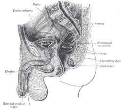 Median sagittal section of male pelvis. (Rectovesical excavation labeled at center right.) | |
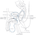
 Male pelvic organs seen from right side. Bladder and rectum distended; relations of peritoneum to the bladder and rectum shown in blue. The arrow points to the rectovesical pouch. | |
| Details | |
| Identifiers | |
| Latin | excavatio rectovesicalis |
| TA98 | A10.1.02.513M |
| TA2 | 3727 |
| FMA | 14727 |
| Anatomical terminology | |
The rectovesical pouch is the pocket that lies between the rectum and the bladder in males in humans and other mammals. It is lined by peritoneum.
Structure
The rectovesical pouch is a space between the rectum and the bladder in men.[1] It lies above the seminal vesicles.[2] It is lined by peritoneum and at its base is the rectoprostatic fascia (Denonvillier's fascia). When a man is upright or supine, it is the lowest part of his peritoneal cavity.[3] It may contain parts of the ileum (lower small intestine) and the sigmoid colon.[2]
In women, the uterus lies between the rectum and the bladder. Therefore, women do not have a rectovesical pouch, but instead have a rectouterine pouch and vesicouterine pouch. After a hysterectomy in women, the remaining peritoneum may be referred to as a rectovesical pouch.[4]
Clinical significance
When a man is upright or supine, the rectovesical pouch is the lowest part of his peritoneal cavity.[3] Because of this, peritoneal fluid and other fluids that enter the peritoneal cavity, including ascites, blood and pus, tend to collect in this pouch.
Additional images
-
 Median sagittal section of pelvis, showing arrangement of fasciae
 -
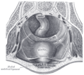 The peritoneum of the male pelvis


See also
References
![]() This article incorporates text in the public domain from page 1152 of the 20th edition of Gray's Anatomy (1918)
This article incorporates text in the public domain from page 1152 of the 20th edition of Gray's Anatomy (1918)
- ^ Friedman, Lana M.; Tsung, James W. (May 2013). "Extending the Focussed Assessment With Sonography for Trauma Examination in Children". 12 (1). Elsevier: 2–17 – via ResearchGate.
{{cite journal}}: Cite journal requires|journal=(help) - ^ a b Jacob, S. (2008). "4 - Abdomen". Human Anatomy. Churchill Livingstone. pp. 71–123. doi:10.1016/B978-0-443-10373-5.50007-5. ISBN 978-0-443-10373-5.
- ^ a b "Colon". Imaging Anatomy: Chest, Abdomen, Pelvis (2nd ed.). Elsevier. 2017. pp. 666–707. doi:10.1016/B978-0-323-47781-9.50032-5. ISBN 978-0-323-47781-9.
- ^ "Ureters and Bladder". Imaging Anatomy: Ultrasound (2nd ed.). Elsevier. 2018. pp. 424–433. doi:10.1016/B978-0-323-54800-7.50047-7. ISBN 978-0-323-54800-7.
This anatomy article is a stub. You can help Wikipedia by expanding it. |