
| Muscle cell | |
|---|---|
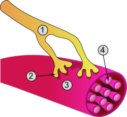 General structure of a skeletal muscle cell and neuromuscular junction: | |
| Details | |
| Location | Muscle |
| Identifiers | |
| Latin | myocytus |
| MeSH | D032342 |
| TH | H2.00.05.0.00002 |
| FMA | 67328 |
| Anatomical terms of microanatomy | |
A muscle cell, also known as a myocyte, is a mature contractile cell in the muscle of an animal.[1] In humans and other vertebrates there are three types: skeletal, smooth, and cardiac (cardiomyocytes).[2] A skeletal muscle cell is long and threadlike with many nuclei and is called a muscle fiber.[3] Muscle cells develop from embryonic precursor cells called myoblasts.[1]
Skeletal muscle cells form by fusion of myoblasts to produce multinucleated cells (syncytia) in a process known as myogenesis.[4][5] Skeletal muscle cells and cardiac muscle cells both contain myofibrils and sarcomeres and form a striated muscle tissue.[6]
Cardiac muscle cells form the cardiac muscle in the walls of the heart chambers, and have a single central nucleus.[7] Cardiac muscle cells are joined to neighboring cells by intercalated discs, and when joined in a visible unit they are described as a cardiac muscle fiber.[8]
Smooth muscle cells control involuntary movements such as the peristalsis contractions in the esophagus and stomach. The Smooth muscle has no myofibrils or sarcomeres and is therefore non-striated. Smooth muscle cells have a single nucleus.
YouTube Encyclopedic
-
1/5Views:6 366 991684 055496 800268 63155 148
-
Muscles, Part 1 - Muscle Cells: Crash Course Anatomy & Physiology #21
-
Anatomy of a muscle cell
-
Muscle Tissues and Sliding Filament Model
-
Anatomy of a skeletal muscle cell | Muscular-skeletal system physiology | NCLEX-RN | Khan Academy
-
Muscle Tissue
Transcription
Romeo and Juliet. Helen and Paris. Tristan and Isolde. These famous star-crossed lovers bring to mind insatiable longing, forbidden love, and tragic separation. And poets and emo rockers love them for it. But you know where else you can find a nice hot romance? Your muscle cells. They’ve got their own famous coupling -- a pretty pair of tiny protein strands called actin and myosin. Romeo and Juliet may have set a chain of tragic events into motion with their infatuation, but deep down in the cells of your muscles, the hot protein action between actin and myosin is actually, literally causing all of your motions. ALL of them. And I don’t just mean voluntary stuff, like walking down the street, or moving your mouth so you can talk or chew chips. Because your muscles also support your weight and help fend off gravity. The amazing thing about your complicated, self-healing, blood-guzzling muscle tissues is that they turn chemical potential energy into mechanical energy, or movement, simply by doing two things -- contracting and relaxing. And that contracting and relaxing is exactly what’s fueled by the constant coupling and separation of biology’s greatest lovers. Somebody get these proteins a movie contract. You will recall from our early lessons on tissues that you’re kept alive and moving by three types of muscle tissue: smooth, cardiac, and skeletal. Your smooth muscle tissue is found in the walls of all your hollow visceral organs, like your stomach, and airways, and blood vessels, where it involuntarily and very usefully pushes fluid and other material around by contracting and relaxing, over and over. Your heart is so important that it gets its very own muscle tissue type -- cardiac muscle, which looks striped, or striated, and also functions involuntarily to keep your blood pumping without you having to think about it. But when you hear the word “muscle,” you probably think of the kind you see on Chris Evans when he first walks out of that machine in Captain America. And those types -- the ones you can see and feel and flex -- are your 640 skeletal muscles. They’re striated like cardiac muscle tissue, but they’re also mostly voluntary, meaning you have to think about using them and activate them with your somatic nervous system. Most of them attach to your skeleton, and create movement by pulling bones in different directions as they contract. Each one of your different skeletal muscles -- like your biceps brachii, or vastus lateralis or gluteus maximus -- is technically its own organ, made up mostly of muscle tissue, but also of connective tissue, blood vessels, and nerve fibers. And because your muscles are voracious energy hogs, each one is rigged up with its own personal nerve to stimulate contraction, and its own artery and vein to keep it well-fed with all the blood, and oxygen, and nutrients it needs to operate. But to understand those operations, we first need to get a grip on the anatomy of a skeletal muscle, which involves fibers within fibers, and lots of layers. Basically, a skeletal muscle is constructed like a really sturdy piece of rope. Thousands of tiny, parallel threads called myofibrils squish together to form muscle fibers, which are your actual muscle cells -- cells with mitochondria, multiple nuclei, and a cellular membrane called a sarcolemma. Those muscle fibers then form larger, string-like bundles called fascicles, which combine to form the larger rope-like muscle organ, like your biceps brachii. Overall, this bundles-of-bundles configuration makes muscle tissue fairly sturdy. But considering how much abuse your muscles take when you do something like pretty simple, like lift a big bag of dog food, it’s no surprise they need a little help. That’s why every muscle contains a few different kinds of supportive sheaths of connective tissue -- the protective reinforcements to keep that bulging muscle from bursting. So that is the structure part of the story. But if you want to get into the nitty-gritty -- the down-and-dirty -- of how you actually move, well, there are rules. Really, just two main rules, and they have to do with proteins. And they’re both true for a lot of the proteins we talk about, whether they’re enzymes or ion channels or receptors or muscle proteins. And these rules are: One. Proteins like to change shape when stuff binds to them. And two. Changing shapes can allow proteins to bind -- or unbind -- with other stuff. So keep those rules in mind, while we see how a muscle fiber contracts and relaxes. Now, remember those tiny myofibrils that bundle up to form your muscle fibers? They’re divided lengthwise into segments called sarcomeres, which contain two even tinier strands of protein -- two different kinds of myofilaments called actin and myosin. And it’s their angsty story of star-crossed love that fuels every movement your body could possibly dream up. A sarcomere contains both thin filaments, made up mostly of two light and twisty actin strands, and thick filaments, composed of thicker, lumpy-looking myosin strands. Each sarcomere is separated by what’s known as a Z line at either end, which is just a border formed by alternating thin filaments in a kind of zig zag pattern. A muscle contracting is all about sarcomeres contracting, bringing those Z-lines closer together. All right, so now comes the romance. When your muscle cells are at rest, your actin & myosin strands don’t touch, but they really, really want to. Specifically, that club-headed myosin wants to get all up-close-and-personal with the actin. When this happens -- and it will, eventually -- it’s called the sliding filament model of muscle contraction. But in the meantime, like in any good love story, the pair have some obstacles to overcome. Namely, actin is blocked by a couple of protein bodyguards -- called tropomyosin and troponin -- which keep getting in the way. Luckily, these guards can be bought off with a little ATP and some calcium. I prefer cash and nachos, but whatever. Remember, ATP is kind of like molecular currency. It contains chemical energy, and your muscles are all about converting chemical energy to motion, so they’re always hungry for more ATP. Your muscle cells have lots of nuclei, but some of them also have a lot of mitochondria, whose sole purpose in life is to crank out ATP. And muscle cells also have their own version of an endoplasmic reticulum -- the cell’s transport and storage system -- but in this case it’s specialized, so it gets a special name: the sarcoplasmic reticulum. Its walls are loaded with calcium pumps -- which use ATP to save up a bunch of calcium ions. And it’s also studded with calcium channels that are linked to voltage-sensitive proteins in the membrane of the muscle cell. Say I want to move my arm. My brain sends an action potential along the motor neuron until it synapses with a muscle cell in my arm. The receptors on that muscle cell are ligand-gated sodium channels, so when the motor neuron releases our old friend acetylcholine into the synapse, the channels open up, and create a rush of sodium into the cell as a graded potential, which, if it’s strong enough, causes nearby voltage-gated sodium channels to open. Now, I want to take a second and point out here that we’re still talking about an action potential, but not in a neuron. This is happening in a muscle cell, people. So that action potential zips along a muscle cell’s membrane, the sarcolemma, which has lots of tubes that run deep inside the cell, called T-Tubules. When the action potential travels down one of those tubes, it eventually triggers the voltage-sensitive proteins that are linked to those calcium channels on the cell’s sarcoplasmic reticulum. When those channels are thrown open, the calcium stored inside rushes into the rest of the cell, and finally myosin is like, YES! Here we go! At this point, the myosin is totally stoked, because the bodyguards that have been frustrating it are in for a big, irresistible distraction. That’s because the protein troponin just loves to bind with calcium, and remember: When stuff binds to proteins, the proteins change shape. So the calcium latches on to the troponin and causes it to pull the other bodyguard protein -- the tropomyosin -- away from the sites on the actin strands that the myosin really wants to get its paws on. And suddenly it’s all, “Okay?” “Okay.” But the only myosin heads that can bind to those newly exposed sites are ones that are ready for action. That is, the ones that have already grabbed a molecule of ATP that’s been floating around, and broken it down into ADP and the leftover phosphate. When a myosin head does that, it moves into an extended position, kinda like a stretched spring -- still holding on to the ADP and phosphate, and still storing the energy that was released when they were broken apart. So after all that, with the myosin primed for action and the bodyguards out of the way, the myosin finally binds to actin, and it is beautiful. When they bind, the myosin releases all that stored energy, and -- in the excitement of it all -- the myosin changes shape. It pulls on its precious actin strand, kind of like pulling a rope hand over fist. In the process, it shrinks the whole sarcomere, and contracts the muscle. That’s the sliding part of the sliding filament model. Now, with its energy spent, that little head has no use for the ADP and the phosphate. So they un-bind with it, because -- remember Rule Number Two, changing shape encourages proteins to bind or unbind with stuff. That unbinding causes a small change in its shape, which lets a fresh ATP binds there in its place. That binding causes another shape change. But this time, it causes the myosin to release from the actin, in a tear-jerking scene like some microscopic re-creation of the finale from Titanic. But fear not! This epic is not quite over! Because this is when the myosin breaks down its new molecule of ATP into ADP and a phosphate, which moves it into the armed position yet again, getting it ready for its next rendezvous. And meanwhile, those calcium pumps are working hard to restock the calcium in the sarcoplasmic reticulum. So they start grabbing the calcium that’s floating around, causing calcium to unbind from the troponin. When it unbinds, the resulting shape-change puts the tropomyosin bodyguards back into place. It’s a circle. Or potentially a big Hollywood franchise. With lots and lots of sequels. It keeps repeating itself many, many times every moment, while I sit here and talk, and while you sit there and eat and text and take notes, the whole drama replaying itself over and over. Kind of like you’ll have to play this video over and over again to get all the little steps of the sliding filament model straightened out. But hey, some stories get better the more you hear them. If you do watch this one again, you will re-learn that your smooth, cardiac, and skeletal muscles create movement by contracting and releasing in a process called the sliding filament model. You’d also re-learn that your skeletal muscles are constructed like a rope made of bundles of protein fibers, and that the smallest strands are your actin and myosin myofilaments. Its their use of calcium and ATP that causes the binding and unbinding that makes sarcomeres contract and relax. Special thanks to our Headmaster of Learning Thomas Frank for his support of Crash Course and free education. And thank you to all of our Patreon patrons who help make Crash Course possible through their monthly contributions. If you like Crash Course and want to help us keep making great new videos like this one, you can check out patreon.com/crashcourse Crash Course is filmed in the Doctor Cheryl C. Kinney Crash Course Studio. This episode was written by Kathleen Yale, edited by Blake de Pastino, and our consultant, is Dr. Brandon Jackson. Our director is Nicholas Jenkins, the editor and script supervisor is Nicole Sweeney, our sound designer is Michael Aranda, and the graphics team is Thought Café.
Structure
The unusual microscopic anatomy of a muscle cell gave rise to its terminology. The cytoplasm in a muscle cell is termed the sarcoplasm; the smooth endoplasmic reticulum of a muscle cell is termed the sarcoplasmic reticulum; and the cell membrane in a muscle cell is termed the sarcolemma.[9] The sarcolemma receives and conducts stimuli.
Skeletal muscle cells

Skeletal muscle cells are the individual contractile cells within a muscle and are more usually known as muscle fibers because of their longer threadlike appearance.[10] A single muscle such as the biceps brachii in a young adult human male contains around 253,000 muscle fibers.[11] Skeletal muscle fibers are the only muscle cells that are multinucleated with the nuclei usually referred to as myonuclei. This occurs during myogenesis with the fusion of myoblasts each contributing a nucleus to the newly formed muscle cell or myotube.[12] Fusion depends on muscle-specific proteins known as fusogens called myomaker and myomerger.[13]
A striated muscle fiber contains myofibrils consisting of long protein chains of myofilaments. There are three types of myofilaments: thin, thick, and elastic that work together to produce a muscle contraction.[14] The thin myofilaments are filaments of mostly actin and the thick filaments are of mostly myosin and they slide over each other to shorten the fiber length in a muscle contraction. The third type of myofilament is an elastic filament composed of titin, a very large protein.
In striations of muscle bands, myosin forms the dark filaments that make up the A band. Thin filaments of actin are the light filaments that make up the I band. The smallest contractile unit in the fiber is called the sarcomere which is a repeating unit within two Z bands. The sarcoplasm also contains glycogen which provides energy to the cell during heightened exercise, and myoglobin, the red pigment that stores oxygen until needed for muscular activity.[14]
The sarcoplasmic reticulum, a specialized type of smooth endoplasmic reticulum, forms a network around each myofibril of the muscle fiber. This network is composed of groupings of two dilated end-sacs called terminal cisternae, and a single T-tubule (transverse tubule), which bores through the cell and emerge on the other side; together these three components form the triads that exist within the network of the sarcoplasmic reticulum, in which each T-tubule has two terminal cisternae on each side of it. The sarcoplasmic reticulum serves as a reservoir for calcium ions, so when an action potential spreads over the T-tubule, it signals the sarcoplasmic reticulum to release calcium ions from the gated membrane channels to stimulate muscle contraction.[14][15]
In skeletal muscle, at the end of each muscle fiber, the outer layer of the sarcolemma combines with tendon fibers at the myotendinous junction.[16][17] Within the muscle fiber pressed against the sarcolemma are multiply flattened nuclei; embryologically, this multinucleate condition results from multiple myoblasts fusing to produce each muscle fiber, where each myoblast contributes one nucleus.[14]
Cardiac muscle cells
The cell membrane of a cardiac muscle cell has several specialized regions, which may include the intercalated disc, and transverse tubules. The cell membrane is covered by a lamina coat which is approximately 50 nm wide. The laminar coat is separable into two layers; the lamina densa and lamina lucida. In between these two layers can be several different types of ions, including calcium.[18]
Cardiac muscle like the skeletal muscle is also striated and the cells contain myofibrils, myofilaments, and sarcomeres as the skeletal muscle cell. The cell membrane is anchored to the cell's cytoskeleton by anchor fibers that are approximately 10 nm wide. These are generally located at the Z lines so that they form grooves and transverse tubules emanate. In cardiac myocytes, this forms a scalloped surface.[18]
The cytoskeleton is what the rest of the cell builds off of and has two primary purposes; the first is to stabilize the topography of the intracellular components and the second is to help control the size and shape of the cell. While the first function is important for biochemical processes, the latter is crucial in defining the surface-to-volume ratio of the cell. This heavily influences the potential electrical properties of excitable cells. Additionally, deviation from the standard shape and size of the cell can have a negative prognostic impact.[18]
Smooth muscle cells
Smooth muscle cells are so-called because they have neither myofibrils nor sarcomeres and therefore no striations. They are found in the walls of hollow organs, including the stomach, intestines, bladder and uterus, in the walls of blood vessels, and in the tracts of the respiratory, urinary, and reproductive systems. In the eyes, the ciliary muscles dilate and contract the iris and alter the shape of the lens. In the skin, smooth muscle cells such as those of the arrector pili cause hair to stand erect in response to cold temperature or fear.[19]
Smooth muscle cells are spindle-shaped with wide middles, and tapering ends. They have a single nucleus and range from 30 to 200 micrometers in length. This is thousands of times shorter than skeletal muscle fibers. The diameter of their cells is also much smaller which removes the need for T-tubules found in striated muscle cells. Although smooth muscle cells lack sarcomeres and myofibrils they do contain large amounts of the contractile proteins actin and myosin. Actin filaments are anchored by dense bodies (similar to the Z discs in sarcomeres) to the sarcolemma.[19]
Development
A myoblast is an embryonic precursor cell that differentiates to give rise to the different muscle cell types.[20] Differentiation is regulated by myogenic regulatory factors, including MyoD, Myf5, myogenin, and MRF4.[21] GATA4 and GATA6 also play a role in myocyte differentiation.[22]
Skeletal muscle fibers are made when myoblasts fuse together; muscle fibers therefore are cells with multiple nuclei, known as myonuclei, with each cell nucleus originating from a single myoblast. The fusion of myoblasts is specific to skeletal muscle, and not cardiac muscle or smooth muscle.
Myoblasts in skeletal muscle that do not form muscle fibers dedifferentiate back into myosatellite cells. These satellite cells remain adjacent to a skeletal muscle fiber, situated between the sarcolemma and the basement membrane[23] of the endomysium (the connective tissue investment that divides the muscle fascicles into individual fibers). To re-activate myogenesis, the satellite cells must be stimulated to differentiate into new fibers.
Myoblasts and their derivatives, including satellite cells, can now be generated in vitro through directed differentiation of pluripotent stem cells.[24]
Kindlin-2 plays a role in developmental elongation during myogenesis.[25]
Function
Muscle contraction in striated muscle
Skeletal muscle contraction
When contracting, thin and thick filaments slide concerning each other by using adenosine triphosphate. This pulls the Z discs closer together in a process called the sliding filament mechanism. The contraction of all the sarcomeres results in the contraction of the whole muscle fiber. This contraction of the myocyte is triggered by the action potential over the cell membrane of the myocyte. The action potential uses transverse tubules to get from the surface to the interior of the myocyte, which is continuous within the cell membrane. Sarcoplasmic reticula are membranous bags that transverse tubules touch but remain separate from. These wrap themselves around each sarcomere and are filled with Ca2+.[26]
Excitation of a myocyte causes depolarization at its synapses, the neuromuscular junctions, which triggers an action potential. With a singular neuromuscular junction, each muscle fiber receives input from just one somatic efferent neuron. Action potential in a somatic efferent neuron causes the release of the neurotransmitter acetylcholine.[27]
When the acetylcholine is released it diffuses across the synapse and binds to a receptor on the sarcolemma, a term unique to muscle cells that refers to the cell membrane. This initiates an impulse that travels across the sarcolemma.[28]
When the action potential reaches the sarcoplasmic reticulum it triggers the release of Ca2+ from the Ca2+ channels. The Ca2+ flows from the sarcoplasmic reticulum into the sarcomere with both of its filaments. This causes the filaments to start sliding and the sarcomeres to become shorter. This requires a large amount of ATP, as it is used in both the attachment and release of every myosin head. Very quickly Ca2+ is actively transported back into the sarcoplasmic reticulum, which blocks the interaction between the thin and thick filament. This in turn causes the muscle cell to relax.[28]
There are four main types of muscle contraction: twitch, treppe, tetanus, and isometric/isotonic. Twitch contraction is the process in which a single stimulus signals a single contraction. In twitch contraction, the length of the contraction may vary depending on the size of the muscle cell. During treppe (or summation) contraction muscles do not start at maximum efficiency; instead, they achieve increased strength of contraction due to repeated stimuli. Tetanus involves a sustained contraction of muscles due to a series of rapid stimuli, which can continue until the muscles fatigue. Isometric contractions are skeletal muscle contractions that do not cause movement of the muscle. However, isotonic contractions are skeletal muscle contractions that do cause movement.[28]
Cardiac muscle contraction
Specialized cardiomyocytes in the sinoatrial node generate electrical impulses that control the heart rate. These electrical impulses coordinate contraction throughout the remaining heart muscle via the electrical conduction system of the heart. Sinoatrial node activity is modulated, in turn, by nerve fibers of both the sympathetic and parasympathetic nervous systems. These systems act to increase and decrease, respectively, the rate of production of electrical impulses by the sinoatrial node.
Evolution
The evolutionary origin of muscle cells in animals is highly debated: One view is that muscle cells evolved once, and thus all muscle cells have a single common ancestor. Another view is that muscles cells evolved more than once, and any morphological or structural similarities are due to convergent evolution, and the development of shared genes that predate the evolution of muscle – even the mesoderm (the mesoderm is the germ layer that gives rise to muscle cells in vertebrates).
Schmid & Seipel (2005)[29] argue that the origin of muscle cells is a monophyletic trait that occurred concurrently with the development of the digestive and nervous systems of all animals, and that this origin can be traced to a single metazoan ancestor in which muscle cells are present. They argue that molecular and morphological similarities between the muscles cells in Cnidaria and Ctenophora are similar enough to those of bilaterians that there would be one ancestor in metazoans from which muscle cells derive. In this case, Schmid & Seipel argue that the last common ancestor of Bilateria, Ctenophora and Cnidaria, was a triploblast (an organism having three germ layers), and that diploblasty, meaning an organism with two germ layers, evolved secondarily, because of their observation of the lack of mesoderm or muscle found in most cnidarians and ctenophores. By comparing the morphology of cnidarians and ctenophores to bilaterians, Schmid & Seipel were able to conclude that there were myoblast-like structures in the tentacles and gut of some species of cnidarians and the tentacles of ctenophores. Since this is a structure unique to muscle cells, these scientists determined based on the data collected by their peers that this is a marker for striated muscles similar to that observed in bilaterians. The authors also remark that the muscle cells found in cnidarians and ctenophores are often contested due to the origin of these muscle cells being the ectoderm rather than the mesoderm or mesendoderm.
The origin of true muscle cells is argued by other authors to be the endoderm portion of the mesoderm and the endoderm. However, Schmid & Seipel (2005)[29] counter skepticism – about whether the muscle cells found in ctenophores and cnidarians are "true" muscle cells – by considering that cnidarians develop through a medusa stage and polyp stage. They note that in the hydrozoans' medusa stage, there is a layer of cells that separate from the distal side of the ectoderm, which forms the striated muscle cells in a way similar to that of the mesoderm; they call this third separated layer of cells the ectocodon. Schmid & Seipel argue that even in bilaterians, not all muscle cells are derived from the mesendoderm: Their key examples are that in both the eye muscles of vertebrates, and the muscles of spiralians, these cells derive from the ectodermal mesoderm, rather than the endodermal mesoderm. Furthermore, they argue that since myogenesis does occur in cnidarians with the help of the same molecular regulatory elements found in the specification of muscle cells in bilaterians, that there is evidence for a single origin for striated muscle.[29]
In contrast to this argument for a single origin of muscle cells, Steinmetz, Kraus, et al. (2012)[30] argue that molecular markers such as the myosin II protein used to determine this single origin of striated muscle predate the formation of muscle cells. They use an example of the contractile elements present in the Porifera, or sponges, that do truly lack this striated muscle containing this protein. Furthermore, Steinmetz, Kraus, et al. present evidence for a polyphyletic origin of striated muscle cell development through their analysis of morphological and molecular markers that are present in bilaterians and absent in cnidarians, ctenophores, and bilaterians. Steinmetz, Kraus, et al. showed that the traditional morphological and regulatory markers such as actin, the ability to couple myosin side chains phosphorylation to higher concentrations of the positive concentrations of calcium, and other MyHC elements are present in all metazoans not just the organisms that have been shown to have muscle cells. Thus, the usage of any of these structural or regulatory elements in determining whether or not the muscle cells of the cnidarians and ctenophores are similar enough to the muscle cells of the bilaterians to confirm a single lineage is questionable according to Steinmetz, Kraus, et al. Furthermore, they explain that the orthologues of the Myc genes that have been used to hypothesize the origin of striated muscle occurred through a gene duplication event that predates the first true muscle cells (meaning striated muscle), and they show that the Myc genes are present in the sponges that have contractile elements but no true muscle cells. Steinmetz, Kraus, et al. also showed that the localization of this duplicated set of genes that serve both the function of facilitating the formation of striated muscle genes, and cell regulation and movement genes, were already separated into striated much and non-muscle MHC. This separation of the duplicated set of genes is shown through the localization of the striated much to the contractile vacuole in sponges, while the non-muscle much was more diffusely expressed during developmental cell shape and change. Steinmetz, Kraus, et al. found a similar pattern of localization in cnidarians except with the cnidarian N. vectensis having this striated muscle marker present in the smooth muscle of the digestive tract. Thus, they argue that the pleisiomorphic trait of the separated orthologues of much cannot be used to determine the monophylogeny of muscle, and additionally argue that the presence of a striated muscle marker in the smooth muscle of this cnidarian shows a fundamental different mechanism of muscle cell development and structure in cnidarians.[30]
Steinmetz, Kraus, et al. (2012)[30] further argue for multiple origins of striated muscle in the metazoans by explaining that a key set of genes used to form the troponin complex for muscle regulation and formation in bilaterians is missing from the cnidarians and ctenophores, and 47 structural and regulatory proteins observed, Steinmetz, Kraus, et al. were not able to find even on unique striated muscle cell protein that was expressed in both cnidarians and bilaterians. Furthermore, the Z-disc seemed to have evolved differently even within bilaterians and there is a great deal of diversity of proteins developed even between this clade, showing a large degree of radiation for muscle cells. Through this divergence of the Z-disc, Steinmetz, Kraus, et al. argue that there are only four common protein components that were present in all bilaterians muscle ancestors and that of these for necessary Z-disc components only an actin protein that they have already argued is an uninformative marker through its pleisiomorphic state is present in cnidarians. Through further molecular marker testing, Steinmetz et al. observe that non-bilaterians lack many regulatory and structural components necessary for bilaterians muscle formation and do not find any unique set of proteins to both bilaterians and cnidarians and ctenophores that are not present in earlier, more primitive animals such as the sponges and amoebozoans. Through this analysis, the authors conclude that due to the lack of elements that bilaterian muscles are dependent on for structure and usage, nonbilaterian muscles must be of a different origin with a different set of regulatory and structural proteins.[30]
In another take on the argument, Andrikou & Arnone (2015)[31] use the newly available data on gene regulatory networks to look at how the hierarchy of genes and morphogens and another mechanism of tissue specification diverge and are similar among early deuterostomes and protostomes. By understanding not only what genes are present in all bilaterians but also the time and place of deployment of these genes, Andrikou & Arnone discuss a deeper understanding of the evolution of myogenesis.[31]
In their paper, Andrikou & Arnone (2015)[31] argue that to truly understand the evolution of muscle cells the function of transcriptional regulators must be understood in the context of other external and internal interactions. Through their analysis, Andrikou & Arnone found that there were conserved orthologues of the gene regulatory network in both invertebrate bilaterians and cnidarians. They argue that having this common, general regulatory circuit allowed for a high degree of divergence from a single well-functioning network. Andrikou & Arnone found that the orthologues of genes found in vertebrates had been changed through different types of structural mutations in the invertebrate deuterostomes and protostomes, and they argue that these structural changes in the genes allowed for a large divergence of muscle function and muscle formation in these species. Andrikou & Arnone were able to recognize not only any difference due to mutation in the genes found in vertebrates and invertebrates but also the integration of species-specific genes that could also cause divergence from the original gene regulatory network function. Thus, although a common muscle patterning system has been determined, they argue that this could be due to a more ancestral gene regulatory network being coopted several times across lineages with additional genes and mutations causing very divergent development of muscles. Thus it seems that the myogenic patterning framework may be an ancestral trait. However, Andrikou & Arnone explain that the basic muscle patterning structure must also be considered in combination with the cis regulatory elements present at different times during development. In contrast with the high level of gene family apparatuses structure, Andrikou and Arnone found that the cis-regulatory elements were not well conserved both in time and place in the network which could show a large degree of divergence in the formation of muscle cells. Through this analysis, it seems that the myogenic GRN is an ancestral GRN with actual changes in myogenic function and structure possibly being linked to later coopts of genes at different times and places.[31]
Evolutionarily, specialized forms of skeletal and cardiac muscles predated the divergence of the vertebrate / arthropod evolutionary line.[32] This indicates that these types of muscle developed in a common ancestor sometime before 700 million years ago (mya). Vertebrate smooth muscle was found to have evolved independently from the skeletal and cardiac muscle types.
Invertebrate muscle cell types
The properties used for distinguishing fast, intermediate, and slow muscle fibers can be different for invertebrate flight and jump muscle.[33] To further complicate this classification scheme, the mitochondrial content, and other morphological properties within a muscle fiber, can change in a tsetse fly with exercise and age.[34]
See also
- List of human cell types derived from the germ layers
- List of distinct cell types in the adult human body
References
- ^ a b Myocytes at the U.S. National Library of Medicine Medical Subject Headings (MeSH)
- ^ Brunet, Thibaut; et al. (2016). "The evolutionary origin of bilaterian smooth and striated myocytes". eLife. 5: 1. doi:10.7554/elife.19607. ISSN 2050-084X. PMC 5167519.
- ^ Saladin, Kenneth S. (2011). Human anatomy (3rd ed.). New York: McGraw-Hill. pp. 72–73. ISBN 9780071222075.
- ^ Scott, W; Stevens, J; Binder-Macleod, SA (2001). "Human skeletal muscle fiber type classifications". Physical Therapy. 81 (11): 1810–1816. doi:10.1093/ptj/81.11.1810. PMID 11694174. Archived from the original on 13 February 2015.
- ^ "Does anyone know why skeletal muscle fibers have peripheral nuclei, but the cardiomyocytes not? What are the functional advantages?". Archived from the original on 19 September 2017.
- ^ Betts, J. Gordon; Young, Kelly A.; Wise, James A.; Johnson, Eddie; Poe, Brandon; Kruse, Dean H.; Korol, Oksana; Johnson, Jody E.; Womble, Mark; Desaix, Peter (6 March 2013). "Cardiac muscle tissue". Retrieved 3 May 2021.
- ^ "Muscle tissues". Archived from the original on 13 October 2015. Retrieved 29 September 2015.
- ^ "Atrial structure, fibers, and conduction" (PDF). Retrieved 5 June 2021.
- ^ Saladin, Kenneth S. (2011). Human anatomy (3rd ed.). New York: McGraw-Hill. pp. 244–246. ISBN 9780071222075.
- ^ "Structure of Skeletal Muscle | SEER Training". training.seer.cancer.gov.
- ^ Klein, CS; Marsh, GD; Petrella, RJ; Rice, CL (July 2003). "Muscle fiber number in the biceps brachii muscle of young and old men". Muscle & Nerve. 28 (1): 62–8. doi:10.1002/mus.10386. PMID 12811774. S2CID 20508198.
- ^ Cho, CH; Lee, KJ; Lee, EH (August 2018). "With the greatest care, stromal interaction molecule (STIM) proteins verify what skeletal muscle is doing". BMB Reports. 51 (8): 378–387. doi:10.5483/bmbrep.2018.51.8.128. PMC 6130827. PMID 29898810.
- ^ Prasad, V; Millay, DP (8 May 2021). "Skeletal muscle fibers count on nuclear numbers for growth". Seminars in Cell & Developmental Biology. 119: 3–10. doi:10.1016/j.semcdb.2021.04.015. PMC 9070318. PMID 33972174. S2CID 234362466.
- ^ a b c d Saladin, K (2012). Anatomy & Physiology: The Unity of Form and Function (6th ed.). New York: McGraw-Hill. pp. 403–405. ISBN 978-0-07-337825-1.
- ^ Sugi, Haruo; Abe, T; Kobayashi, T; Chaen, S; Ohnuki, Y; Saeki, Y; Sugiura, S; Guerrero-Hernandez, Agustin (2013). "Enhancement of force generated by individual myosin heads in skinned rabbit psoas muscle fibers at low ionic strength". PLOS ONE. 8 (5): e63658. Bibcode:2013PLoSO...863658S. doi:10.1371/journal.pone.0063658. PMC 3655179. PMID 23691080.
- ^ Charvet, B; Ruggiero, F; Le Guellec, D (April 2012). "The development of the myotendinous junction. A review". Muscles, Ligaments and Tendons Journal. 2 (2): 53–63. PMC 3666507. PMID 23738275.
- ^ Bentzinger, CF; Wang, YX; Rudnicki, MA (1 February 2012). "Building muscle: molecular regulation of myogenesis". Cold Spring Harbor Perspectives in Biology. 4 (2): a008342. doi:10.1101/cshperspect.a008342. PMC 3281568. PMID 22300977.
- ^ a b c Ferrari, Roberto. "Healthy versus sick myocytes: metabolism, structure and function" (PDF). oxfordjournals.org/en. Oxford University Press. Archived from the original (PDF) on 19 February 2015. Retrieved 12 February 2015.
- ^ a b Betts, J. Gordon; Young, Kelly A.; Wise, James A.; Johnson, Eddie; Poe, Brandon; Kruse, Dean H.; Korol, Oksana; Johnson, Jody E.; Womble, Mark; Desaix, Peter (6 March 2013). "Smooth muscle". Retrieved 10 June 2021.
- ^ page 395, Biology, Fifth Edition, Campbell, 1999
- ^ Perry R, Rudnick M (2000). "Molecular mechanisms regulating myogenic determination and differentiation". Front Biosci. 5: D750–67. doi:10.2741/Perry. PMID 10966875.
- ^ Zhao R, Watt AJ, Battle MA, Li J, Bandow BJ, Duncan SA (May 2008). "Loss of both GATA4 and GATA6 blocks cardiac myocyte differentiation and results in acardia in mice". Dev. Biol. 317 (2): 614–9. doi:10.1016/j.ydbio.2008.03.013. PMC 2423416. PMID 18400219.
- ^ Zammit, PS; Partridge, TA; Yablonka-Reuveni, Z (November 2006). "The skeletal muscle satellite cell: the stem cell that came in from the cold". Journal of Histochemistry and Cytochemistry. 54 (11): 1177–91. doi:10.1369/jhc.6r6995.2006. PMID 16899758.
- ^ Chal J, Oginuma M, Al Tanoury Z, Gobert B, Sumara O, Hick A, Bousson F, Zidouni Y, Mursch C, Moncuquet P, Tassy O, Vincent S, Miyazaki A, Bera A, Garnier JM, Guevara G, Heston M, Kennedy L, Hayashi S, Drayton B, Cherrier T, Gayraud-Morel B, Gussoni E, Relaix F, Tajbakhsh S, Pourquié O (August 2015). "Differentiation of pluripotent stem cells to muscle fiber to model Duchenne muscular dystrophy". Nature Biotechnology. 33 (9): 962–9. doi:10.1038/nbt.3297. PMID 26237517. S2CID 21241434.

- ^ Dowling JJ, Vreede AP, Kim S, Golden J, Feldman EL (2008). "Kindlin-2 is required for myocyte elongation and is essential for myogenesis". BMC Cell Biol. 9: 36. doi:10.1186/1471-2121-9-36. PMC 2478659. PMID 18611274.
- ^ "Structure, and Function of Skeletal Muscles". courses.washington.edu. Archived from the original on 15 February 2015. Retrieved 13 February 2015.
- ^ "Muscle Fiber Excitation". courses.washington.edu. University of Washington. Archived from the original on 27 February 2015. Retrieved 11 February 2015.
- ^ a b c Ziser, Stephen. "Muscle Cell Anatomy & Function" (PDF). www.austincc.edu. Archived (PDF) from the original on 23 September 2015. Retrieved 12 February 2015.
- ^ a b c Seipel, Katja; Schmid, Volker (1 June 2005). "Evolution of striated muscle: Jellyfish and the origin of triploblasty". Developmental Biology. 282 (1): 14–26. doi:10.1016/j.ydbio.2005.03.032. PMID 15936326.
- ^ a b c d Steinmetz, Patrick R.H.; Kraus, Johanna E.M.; Larroux, Claire; Hammel, Jörg U.; Amon-Hassenzahl, Annette; Houliston, Evelyn; et al. (2012). "Independent evolution of striated muscles in cnidarians and bilaterians". Nature. 487 (7406): 231–234. Bibcode:2012Natur.487..231S. doi:10.1038/nature11180. PMC 3398149. PMID 22763458.
- ^ a b c d Andrikou, Carmen; Arnone, Maria Ina (1 May 2015). "Too many ways to make a muscle: Evolution of GRNs governing myogenesis". Zoologischer Anzeiger. Special Issue: Proceedings of the 3rd International Congress on Invertebrate Morphology. 256: 2–13. doi:10.1016/j.jcz.2015.03.005.
- ^ OOta, S.; Saitou, N. (1999). "Phylogenetic relationship of muscle tissues deduced from the superimposition of gene trees". Molecular Biology and Evolution. 16 (6): 856–867. doi:10.1093/oxfordjournals.molbev.a026170. ISSN 0737-4038. PMID 10368962.
- ^ Hoyle, Graham (1983). "8. Muscle cell diversity". Muscles and Their Neural Control. New York, NY: John Wiley & Sons. pp. 293–299. ISBN 9780471877097.
- ^ Anderson, M.; Finlayson, L.H. (1976). "The effect of exercise on the growth of mitochondria and myofibrils in the flight muscles of the Tsetse fly, Glossina morsitans". J. Morphol. 150 (2): 321–326. doi:10.1002/jmor.1051500205. S2CID 85719905.
External links
 Media related to Myocytes at Wikimedia Commons
- Structure of a Muscle Cell