
| Flexor digitorum superficialis | |
|---|---|
 | |
 Flexor digitorum superficialis | |
| Details | |
| Origin | Medial epicondyle of the humerus (common flexor tendon) as well as parts of the radius and ulna |
| Insertion | Anterior margins on the base of the middle phalanges of the four fingers |
| Artery | Radial artery |
| Nerve | Median nerve |
| Actions | Flexor of fingers (primarily at proximal interphalangeal joints) |
| Antagonist | Extensor digitorum muscle |
| Identifiers | |
| Latin | musculus flexor digitorum superficialis |
| TA98 | A04.6.02.033 |
| TA2 | 2486 |
| FMA | 38469 |
| Anatomical terms of muscle | |
Flexor digitorum superficialis (flexor digitorum sublimis) is an extrinsic flexor muscle of the fingers at the proximal interphalangeal joints.
It is in the anterior compartment of the forearm. It is sometimes considered to be the deepest part of the superficial layer of this compartment,[1][2] and sometimes considered to be a distinct, "intermediate layer" of this compartment.[3] It is relatively common for the Flexor digitorum superficialis to be missing from the little finger, bilaterally and unilaterally, which can cause problems when diagnosing a little finger injury.[4]
YouTube Encyclopedic
-
1/3Views:3 23420 67827 593
-
Arm Muscles: 26 Flexor Digitorum Superficialis
-
flexor digitorum superficialis
-
Flexor digitorum profundus muscle - Origin, Insertion, Innervation & Function - Anatomy | Kenhub
Transcription
Structure
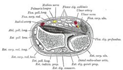
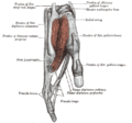
The muscle has two classically described heads – the humeroulnar and radial – and it is between these heads that the median nerve and ulnar artery pass. The ulnar collateral ligament of elbow joint gives its origin to part of this muscle.
Four long tendons come off this muscle near the wrist and travel through the carpal tunnel formed by the flexor retinaculum. These tendons, along with those of flexor digitorum profundus, are enclosed by a common flexor sheath. The tendons attach to the anterior margins on the bases of the intermediate phalanges of the four fingers. These tendons have a split (Camper's Chiasm) at the end of them through which the tendons of flexor digitorum profundus pass.
Innervation
The Flexor digitorium superficialis muscle is innervated by the median nerve (C7, C8, T1).[5]
Function
The primary function of flexor digitorum superficialis is flexion of the middle phalanges of the four fingers (excluding the thumb) at the proximal interphalangeal joints, however under continued action it also flexes the metacarpophalangeal joints and wrist joint.
To test flexor digitorum superficialis, one finger is flexed at the proximal interphalangeal joint against resistance, while the remaining three fingers are held fully extended (to inactivate flexor digitorum profundus).
Additional images
-
 Tendons of forefinger and vincula tendina.
 -
 Cross-section through the middle of the forearm.
 -
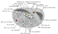 Transverse section across the wrist and digits.
 -
 The mucous sheaths of the tendons on the front of the wrist and digits.
 -
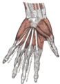 The muscles of the left hand. Palmar surface.
 -
 The radial and ulnar arteries.
 -

-
 Flexor digitorum superficialis muscle
 -
 Flexor digitorum superficialis muscle
 -
 Flexor digitorum superficialis muscle
 -
 Flexor digitorum superficialis muscle
 -
 Flexor digitorum superficialis muscle
 -
 Muscles of upper limb. Cross section.


References
- ^ MedicalMnemonics.com: 273 1117
- ^ "Dissector Answers – Forearm & Wrist". Archived from the original on 2011-07-18. Retrieved 2008-01-17.
- ^ "uams.edu". Archived from the original on 2008-01-19. Retrieved 2008-01-17.
- ^ Townley, W. A.; Swan, M. C.; Dunn, R. L. R. (June 2010). "J Hand Surg Eur". Journal of Hand Surgery (European Volume). 35 (5): 417–418. doi:10.1177/1753193409358523. PMID 20515987.
- ^ Lutsky KF, Giang EL, Matzon JL (January 2015). "Flexor tendon injury, repair and rehabilitation". Orthopedic Clinics of North America. 46 (1): 67–76. doi:10.1016/j.ocl.2014.09.004. PMID 25435036.
External links
- Illustration: upper-body/flexor-digitorum-superficialis from The Department of Radiology at the University of Washington