
| Teres minor muscle | |
|---|---|
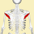
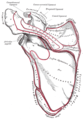
 Teres minor muscle (shown in red), seen from behind. | |
 Muscles on the dorsum of the left scapula, and the Triceps brachii muscle: #3 is Latissimus dorsi muscle #5 is Teres major muscle #6 is Teres minor muscle #7 is Supraspinatus muscle #8 is Infraspinatus muscle #13 is long head of Triceps brachii muscle | |
| Details | |
| Origin | Lateral border of the scapula |
| Insertion | Inferior facet of greater tubercle of the humerus |
| Artery | Posterior circumflex humeral artery and the circumflex scapular artery |
| Nerve | Axillary nerve (C5-C6) |
| Actions | Laterally rotates the arm, stabilizes humerus |
| Identifiers | |
| Latin | musculus teres minor |
| TA98 | A04.6.02.010 |
| TA2 | 2459 |
| FMA | 32550 |
| Anatomical terms of muscle | |
The teres minor (Latin teres meaning 'rounded') is a narrow, elongated muscle of the rotator cuff. The muscle originates from the lateral border and adjacent posterior surface of the corresponding right or left scapula and inserts at both the greater tubercle of the humerus and the posterior surface of the joint capsule.[1]
The primary function of the teres minor is to modulate the action of the deltoid, preventing the humeral head from sliding upward as the arm is abducted. It also functions to rotate the humerus laterally. The teres minor is innervated by the axillary nerve.[2]
YouTube Encyclopedic
-
1/5Views:44 51345 60533 83715 9411 392
-
Teres Minor Muscle - Origin, Insertion, Innervation & Action - Human Anatomy | Kenhub
-
Teres Minor, Why It Is Important - Everything You Need To Know - Dr. Nabil Ebraheim
-
Teres Minor - AnatomyOnlineCourse
-
Treating Trigger Points - Teres Minor
-
Teres Minor Muscle Anatomy
Transcription
Hello again, everyone! It’s Matt from Kenhub, and in this tutorial, we will discuss the origin, insertion, innervation, and action of the teres minor muscle. Along with the supraspinatus muscle, infraspinatus muscle, and subscapularis muscle, the teres minor makes up the rotator cuff, which is a functional, anatomical unit in the upper arm. On this image, we can see the teres minor highlighted in green. All the muscles of the rotator cuff originate from the scapula and insert in the humerus, but the teres minor muscle specifically originates from the lateral scapula border and inserts on the greater tubercle of the humerus. In terms of innervation, the axillary nerve seen on this image, highlighted in green, will innervate the teres minor. The axillary nerve originates from the posterior cord of the brachial plexus at the level of your armpit. The rotator cuff, as the name suggests, plays a major role in the internal and external rotation of the upper arm and the shoulder joint. Its main function is to stabilize the glenoid cavity and keep the humeral head centered in the joint socket. This joint is the most flexible in the human body, and this group of muscles tighten around the joint to prevent a pinch during shoulder movement. The teres minor muscle’s function consists primarily of external rotation, partly retroversion and adduction as well. It works in conjunction with the infraspinatus muscle performing many of the same actions. Because they share a first name, it’s easy to confuse the function and anatomy of the teres major and the teres minor. One memory trigger is to think of the teres major as performing the same action of a major muscle (its big brother), the latissimus dorsi, and the teres minor as performing the same action of a minor muscle, the infraspinatus. This video is more fun than reading a textbook, right? If you want more videos, interactive quizzes, articles, and an atlas of human anatomy, click on the “Take me to Kenhub” button. It is time to say goodbye to your old textbooks and say hello to your new anatomy learning partner, Kenhub! See you there! https://www.kenhub.com
Structure
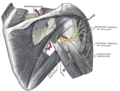
It arises from the dorsal surface of the axillary border of the scapula for the upper two-thirds of its extent, and from two aponeurotic laminae, one of which separates it from the infraspinatus muscle, the other from the teres major muscle.
Its fibers run obliquely upwards and laterally; the upper ones end in a tendon which is inserted into the lowest of the three impressions on the greater tubercle of the humerus; the lowest fibers are inserted directly into the humerus immediately below this impression.
Relations
The teres minor originates at the lateral border and adjacent posterior surface of the scapula. It inserts at the greater tubercle of the humerus. The tendon of this muscle passes across, and is united with, the posterior part of the capsule of the shoulder-joint.
Innervation
The muscle is innervated by the posterior branch of axillary nerve where it forms a pseudoganglion.[3] A pseudoganglion has no nerve cells but nerve fibres are present. Damage to the fibers innervating the teres minor is clinically significant.
Variation
Sometimes a group of muscle fibres from teres minor may be fused with infraspinatus.
Function
The infraspinatus and teres minor attach to head of the humerus; as part of the rotator cuff they help hold the humeral head in the glenoid cavity of the scapula. They work in tandem with the posterior deltoid to externally (laterally) rotate the humerus, as well as adduction. Teres Minor can produce only very small scapular plane adduction during maximal contraction (Hughes RE, An KN 1996) with adductor moment arm of approximately 0.2 cm at 45° of shoulder internal rotation and approximately 0.1 cm at 45° of shoulder external rotation.
Clinical significance
Injury
There are two types of rotator cuff injuries: acute tears and chronic tears. Acute tears occur as a result of a sudden movement. This might include throwing a powerful pitch, holding a fast moving rope during water sports, falling over onto an outstretched hand at speed, or making a sudden thrust with the paddle in kayaking. A chronic tear develops over a period of time. They usually occur at or near the tendon, as a result of the tendon rubbing against the underlying bone.[4] The teres minor is typically normal following a rotator cuff tear.[5]
Imaging


Atrophy of the teres minor muscle is often a consequence of a rotator cuff tear, but common isolated teres minor atrophies have also been found. A quadrangular space syndrome causes excessive and or chronically compression of the structures which pass through this anatomical tunnel. The axillary nerve and the posterior humeral circumflex artery pass through the space. People affected note shoulder pain and paresthesia down the arm first and foremost in abduction, extension, external rotation and overhead activity. Selective atrophy of the teres minor muscle has been seen and pulled together directly with compression of the corresponding axillary nerve branch or posterior humeral circumflex artery. Fibrous bands, cysts of the glenoid labrum, lipoma or dilated veins can occupy the quadrilateral space pathologically. Similar symptoms are common with anterior shoulder dislocation, humeral neck fracture, brachial plexus injury and thoracic outlet and inlet syndrome. It is important to include those pathologies for a complete as possible differential diagnosis.
Ultrasonography is a tool to detect a fatty degenerative atrophy of the teres minor and shows in affected muscles increased echogenicity and betimes a slight reduction in muscle bulk. MR imaging helps to consolidate the diagnosis of neurogenic muscle atrophy. Extracellular edema after traumatic events causing neural damage show an increased signal intensity on T2-weighted MRI sequences and normal intensity on T1-weighted sequences. Posterior humeral circumflex artery compression and reduced blood flow in stressful arm positions and or maneuvers can be diagnosed by a Doppler ultrasonography. The nerve should be detected adjacent to the vessel. In an elevated arm position the axillary neurovascular bundle can be seen at the posterior axillary fold just before it perforates the deltoideus, while the posterior course is well visible in the neutral position. For a detailed assessment of the artery, a MR angiography is required. The major task of an ultrasonographic examination is to rule out any space occupying mass. Additional electromyography is helpful to reveal any decelerated nerve conduction velocity, and thus denervation of the concerned muscle.[6]
Additional images
-
 Position of the teres minor muscles (shown in red). Animation.
 -
 Suprascapular and axillary nerves of right side, seen from behind. (Teres minor is visible at center.)
 -
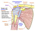 Diagram of the human shoulder joint, front view
 -
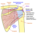 Diagram of the human shoulder joint, back view
 -
 Left scapula. Dorsal surface.
 -
 Left humerus. Posterior view.
 -
 The scapular and circumflex arteries.
 -
 The suprascapular, axillary, and radial nerves.
 -
 Teres minor muscle


See also
References
![]() This article incorporates text in the public domain from page 441 of the 20th edition of Gray's Anatomy (1918)
This article incorporates text in the public domain from page 441 of the 20th edition of Gray's Anatomy (1918)
- ^ Saladin, Kenneth (2015). Anatomy & Physiology: The Unity of Form and Function (7 ed.). New York: McGraw-Hill Education. p. 345. ISBN 9780073403717.
- ^ Saladin, Kenneth (2015). Anatomy & Physiology: The Unity of Form and Function (7 ed.). New York: McGraw-Hill Education. p. 345. ISBN 9780073403717.
- ^ Gitlin, G (October 1957). "Concerning the gangliform enlargement (pseudoganglion) on the nerve to the teres minor muscle". Journal of Anatomy. 91 (4): 466–70. PMC 1244902. PMID 13475146.
- ^ Bahr, Ronald. Ed. Clinical Guide to Sports Injuries. Gazette bok. ISBN 0-7360-4117-6.
- ^ Melis, Barbara; DeFranco, Michael; Ladermann, Alexandre; Barthelemy, Renaud; Walch, Gilles (2011). "The teres minor muscle in rotator cuff tendon tears". Skeletal Radiology. 40 (10): 1335–1344. doi:10.1007/s00256-011-1178-3. PMID 21604212. S2CID 8639793. Retrieved 28 November 2016.
- ^ Brestas, P.S.; et al. (September 2006). ". Ultrasound findings of teres minor denervation in suspected quadrilateral space syndrome". J Clin Ultrasound. 34 (7): 343–7. doi:10.1002/jcu.20239. PMID 16869012. S2CID 6341877.
External links
- Anatomy figure: 03:03-05 at Human Anatomy Online, SUNY Downstate Medical Center
- ExRx