

Tamaño de esta previsualización: 800 × 342 píxeles. Otras resoluciones: 320 × 137 píxeles · 640 × 274 píxeles · 1276 × 546 píxeles.
Ver la imagen en su resolución original (1276 × 546 píxeles; tamaño de archivo: 539 kB; tipo MIME: image/png)
Resumen
| Descripción |
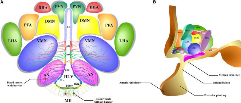
English: A schematic representation of the hypothalamic nuclei and the distribution of tanycytes over the wall of the third ventricle (III-V). (A) Coronal view of the approximate location of the hypothalamic nuclei and tanycytes. Ciliated ependymocytes (ep) line the dorsal wall of the III-V. The α1d-tanycytes (α1d) and α1v-tanycytes (α1v) have long projections that make contact with the neurons of the VMN. α2-tancycytes (α2) have projections to the AN and blood vessels. In a more ventral section of the III-V, the β1d-tanycytes (β1d) and β1v-tanycytes (β1v) make projections to the AN, making contact with orexigenic and anorexigenic neurons and blood vessels. In the floor of the III-V, the β2la-tanycytes (β2la) and β2me-tanycytes (β2me) are joined by tight junctions forming part of the median eminence (ME)-cerebrospinal fluid (CSF) barrier, and their projections make contact with the fenestrated blood vessels of the ME. (B) Sagittal view of the distribution of the hypothalamic nuclei. Ep: ependymocytes; AN: arcuate nucleus; VMN: ventromedial nucleus; DMN: dorsomedial nucleus; PVN: periventricular nucleus; DHA: dorsal hypothalamic area; PFA: perifornical area; LHA: lateral hypothalamic area; SCN: suprachiasmatic nucleus; SON: supraoptic nucleus; POA: preoptic area; MB: mammillary bodies; ME: median eminence; III-V: third ventricle. |
| Fecha | |
| Fuente | Elizondo-Vega R, Cortes-Campos C, Barahona MJ, Oyarce KA, Carril CA, García-Robles MA. The role of tanycytes in hypothalamic glucosensing. Journal of Cellular and Molecular Medicine. 2015;19(7):1471-1482. doi:10.1111/jcmm.12590. https://www.ncbi.nlm.nih.gov/pmc/articles/PMC4511346/ |
| Autor | Roberto Elizondo-Vega, Christian Cortes-Campos, Maria J Barahona, Karina A Oyarce, Claudio A Carril, and Maria A García-Robles |

Licencia
Este archivo está disponible bajo la licencia Creative Commons Atribución 4.0 Internacional.
- Eres libre:
- de compartir – de copiar, distribuir y transmitir el trabajo
- de remezclar – de adaptar el trabajo
- Bajo las siguientes condiciones:
- atribución – Debes otorgar el crédito correspondiente, proporcionar un enlace a la licencia e indicar si realizaste algún cambio. Puedes hacerlo de cualquier manera razonable pero no de manera que sugiera que el licenciante te respalda a ti o al uso que hagas del trabajo.
Historial del archivo
Haz clic sobre una fecha y hora para ver el archivo tal como apareció en ese momento.
| Fecha y hora | Miniatura | Dimensiones | Usuario | Comentario | |
|---|---|---|---|---|---|
| actual | 15:41 15 sep 2018 | | 1276 × 546 (539 kB) | Was a bee | {{Information |Description={{en|1=A schematic representation of the hypothalamic nuclei and the distribution of tanycytes over the wall of the third ventricle (III-V). (A) Coronal view of the approximate location of the hypothalamic nuclei and tanycytes. Ciliated ependymocytes (ep) line the dorsal wall of the III-V. The α1d-tanycytes (α1d) and α1v-tanycytes (α1v) have long projections that make contact with the neurons of the VMN. α2-tancycytes (α2) have projections to the AN and blood vessel... |
Usos del archivo
Las siguientes páginas usan este archivo:
Uso global del archivo
Las wikis siguientes utilizan este archivo:
- Uso en ar.wikipedia.org
- Uso en de.wikipedia.org
- Uso en fr.wikibooks.org