

Ver la imagen en su resolución original ((Imagen SVG, nominalmente 1142 × 1567 pixels, tamaño de archivo: 1,44 MB))
Resumen
| Descripción |
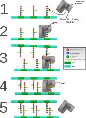
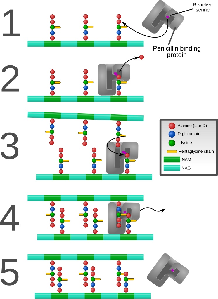
English: Diagram depicting formation of cross-links in the bacterial cell wall by a penicillin binding protein (PBP, an enzyme).
1. The bacterial cell wall consists of strands of repeating N-acetylglucosamine (NAG) and N-acetylmuramic acid (NAM) subunits. The NAM subunits have short peptide chains attached to them. (The exact composition of these can vary. The proximal alanine is usually L-ala and the distal two are usually D-ala.) These chains, in turn, are bound to chains of 5 glycine residues that will be used in cross-linking. 2. The PBP forms a bond with the peptide side chain at the second most distal alanine residue. This displaces the most distal alanine residue. 3. Another strand of bacterial cell wall arrives. The free end of one of the pentaglycine chains displaces the PBP and forms a bond with the terminal alanine on the other strand. 4. After being displaced, the PBP diffuses away. 5. The formation of one cross-link is complete. |
| Fecha | |
| Fuente | Trabajo propio |
| Autor | Mcstrother |
| Otras versiones |
[]
|


Licencia
- Eres libre:
- de compartir – de copiar, distribuir y transmitir el trabajo
- de remezclar – de adaptar el trabajo
- Bajo las siguientes condiciones:
- atribución – Debes otorgar el crédito correspondiente, proporcionar un enlace a la licencia e indicar si realizaste algún cambio. Puedes hacerlo de cualquier manera razonable pero no de manera que sugiera que el licenciante te respalda a ti o al uso que hagas del trabajo.
Historial del archivo
Haz clic sobre una fecha y hora para ver el archivo tal como apareció en ese momento.
| Fecha y hora | Miniatura | Dimensiones | Usuario | Comentario | |
|---|---|---|---|---|---|
| actual | 19:26 9 sep 2011 | | 1142 × 1567 (1,44 MB) | <bdi>Mcstrother</bdi> | Major revision. Corrected inaccuracies in previous image. |
| 04:15 3 may 2011 |  | 1139 × 1062 (850 kB) | <bdi>Mcstrother</bdi> | Changed all fonts to Liberation Sans | |
| 03:46 10 abr 2011 |  | 1139 × 1062 (850 kB) | <bdi>Mcstrother</bdi> | Changed color of carbohydrate chain. | |
| 03:30 7 mar 2011 |  | 1139 × 1062 (835 kB) | <bdi>Mcstrother</bdi> | {{Information |Description ={{en|1=Diagram depicting formation of cross-links in the bacterial cell wall by a penicillin binding protein (PBP, an enzyme). 1. The bacterial cell wall consists of strands of repeating N-acetylglucosamine (NAG) and N-ace |
Usos del archivo
Las siguientes páginas usan este archivo:
Uso global del archivo
Las wikis siguientes utilizan este archivo:
- Uso en en.wikipedia.org
- Uso en fa.wikipedia.org
- Uso en ga.wikipedia.org
- Uso en gl.wikipedia.org
- Uso en hu.wikipedia.org
- Uso en it.wikipedia.org
- Uso en mk.wikipedia.org
- Uso en th.wikipedia.org