
Gray760.png (500 × 323 píxeles; tamaño de archivo: 23 kB; tipo MIME: image/png)
Resumen
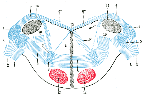
Terminal nuclei of the cochlear nerve, with their upper connections. (Schematic.) The vestibular nerve with its terminal nuclei and their efferent fibers have been suppressed. On the other hand, in order not to obscure the trapezoid body, the efferent fibers of the terminal nuclei on the right side have been resected in a considerable portion of their extent. The trapezoid body, therefore, shows only one-half of its fibers, viz., those which come from the left.
1. Vestibular nerve, divided at its entrance into the medulla oblongata.
2. Cochlear nerve.
3. Accessory nucleus of acoustic nerve.
4. Tuberculum acusticum.
5. Efferent fibers of accessory nucleus.
6. Efferent fibers of tuberculum acusticum, forming the striae medullares, with 6’, their direct bundle going to the superior olivary nucleus of the same side; 6’’, their decussating bundles going to the superior olivary nucleus of the opposite side.
7. Superior olivary nucleus.
8. Trapezoid body.
9. Trapezoid nucleus.
10. Central acoustic tract (lateral lemniscus).
11. Raphé.
12. Cerebrospinal fasciculus.
13. Fourth ventricle.
14. Inferior peduncle.
| Descripción | Terminal nuclei of the cochlear nerve, with their upper connections. (Schematic.) The vestibular nerve with its terminal nuclei and their efferent fibers have been suppressed. On the other hand, in order not to obscure the trapezoid body, the efferent fibers of the terminal nuclei on the right side have been resected in a considerable portion of their extent. The trapezoid body, therefore, shows only one-half of its fibers, viz., those which come from the left. 1. Vestibular nerve, divided at its entrance into the medulla oblongata. 2. Cochlear nerve. 3. Accessory nucleus of acoustic nerve. 4. Tuberculum acusticum. 5. Efferent fibers of accessory nucleus. 6. Efferent fibers of tuberculum acusticum, forming the striae medullares, with 6’, their direct bundle going to the superior olivary nucleus of the same side; 6’’, their decussating bundles going to the superior olivary nucleus of the opposite side. 7. Superior olivary nucleus. 8. Trapezoid body. 9. Trapezoid nucleus. 10. Central acoustic tract (lateral lemniscus). 11. Raphé. 12. Cerebrospinal fasciculus. 13. Fourth ventricle. 14. Inferior peduncle. (Testut.) | ||||||||||||||||||||
| Plate | 760 | ||||||||||||||||||||
| Fecha | antes de 1858 | ||||||||||||||||||||
| Fuente |
|
||||||||||||||||||||
| Autor |
|
||||||||||||||||||||
.jpg)
Libro
| Henry Gray: Gray's Anatomy
|
|||||||||||||||||||||||
|---|---|---|---|---|---|---|---|---|---|---|---|---|---|---|---|---|---|---|---|---|---|---|---|
| Autor |
|
-_Title_page.png) | |||||||||||||||||||||
| Director de edición |
Revised by Warren H. Lewis |
||||||||||||||||||||||
| Ilustrador |
|
||||||||||||||||||||||
| Título | |||||||||||||||||||||||
| Edición |
20 |
||||||||||||||||||||||
| Editorial | |||||||||||||||||||||||
| Object type |
edición, traducción o versión |
||||||||||||||||||||||
| Visión general | list of all the plates | ||||||||||||||||||||||
| Idioma |
inglés |
||||||||||||||||||||||
| Fecha de publicación |
1918 |
||||||||||||||||||||||
| Lugar de publicación |
Filadelfia / Nueva York |
||||||||||||||||||||||
| Fuente | Bartleby | ||||||||||||||||||||||

Licencia
Este archivo está en el dominio público porque es una exploración mecánica simple o fotocopia de un original en dominio público, o (con las pruebas disponibles) es tan similar a un documento escaneado o fotocopia que no se puede aplicar protección de derechos de autor. También puede suceder que los derechos de autor de esta imagen hayan expirado debido a la fecha de publicación o la muerte del autor (si es posible, añadirla aparte). El propio contenido original se encuentra en dominio público por las siguientes razones:
Esta etiqueta está diseñada para usarse cuando sea necesario afirmar que las mejoras (por ejemplo, brillo, contraste, juego de color, nitidez) no son de por sí suficientemente creativas para generar un nuevo derecho de autor. Se puede utilizar tanto cuando no se sabe si se han hecho mejoras como cuando las mejoras son claras, pero insuficientes. Para las imágenes primitivas sin contraste puede utilizar la plantilla {{PD-old}} adecuadamente insertada. Para utilizarla, véase Commons:Cuándo usar la etiqueta PD-scan.  | ||||
Historial del archivo
Haz clic sobre una fecha y hora para ver el archivo tal como apareció en ese momento.
| Fecha y hora | Miniatura | Dimensiones | Usuario | Comentario | |
|---|---|---|---|---|---|
| actual | 20:52 23 ene 2007 | | 500 × 323 (23 kB) | Pngbot | optimized with optipng |
| 05:36 29 ene 2006 |  | 500 × 323 (39 kB) | Arcadian | {{Gray's Anatomy plate}} |
Usos del archivo
La siguiente página usa este archivo:
Uso global del archivo
Las wikis siguientes utilizan este archivo:
- Uso en ar.wikipedia.org
- Uso en bg.wikipedia.org
- Uso en de.wikipedia.org
- Uso en de.wikibooks.org
- Uso en en.wikipedia.org
- Uso en fa.wikipedia.org
- Uso en ja.wikipedia.org
- Uso en kk.wikipedia.org
- Uso en nl.wikipedia.org
- Uso en pl.wikipedia.org
- Uso en zh.wikipedia.org