
| Septum transversum | |
|---|---|
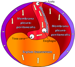 Diaphragm of embryo. | |
 | |
| Details | |
| Carnegie stage | 10 |
| Precursor | mesenchyme |
| Gives rise to | diaphragm / Ventral mesentery |
| Identifiers | |
| TE | transversum_by_E5.2.0.4.0.0.2 E5.2.0.4.0.0.2 |
| Anatomical terminology | |
The septum transversum is a thick mass of cranial mesenchyme, formed in the embryo, that gives rise to parts of the thoracic diaphragm and the ventral mesentery of the foregut in the developed human being and other mammals.
YouTube Encyclopedic
-
1/3Views:19 7562 7705 717
-
Heart Tubes - Embryology
-
Dr.Sherif fahmy -Embryology 19 - " Glands- Muscular system- Septum transversum" ,,
-
Dr Sherif embryology 20 (Development of diaphragm & skull)
Transcription
Origins
The septum transversum originally arises as the most cranial part of the mesenchyme on day 22.[1] During craniocaudal folding, it assumes a position cranial to the developing heart at the level of the cervical vertebrae.[1] During subsequent weeks the dorsal end of the embryo grows much faster than its ventral counterpart resulting in an apparent descent of the ventrally located septum transversum. At week 8, it can be found at the level of the thoracic vertebrae.[1][2]
Nerve supply
After successful craniocaudal folding the septum transversum picks up innervation from the adjacent ventral rami of spinal nerves C3, C4 and C5, thus forming the precursor of the phrenic nerve. During the descent of the septum, the phrenic nerve is carried along and assumes its descending pathway.
During embryonic development of the thoracic diaphragm, myoblast cells from the septum invade the other components of the diaphragm. They thus give rise to the motor and sensory innervation of the muscular diaphragm by the phrenic nerve.
Derivatives
The cranial part of the septum transversum gives rise to the central tendon of the diaphragm,[1] and is the origin of the myoblasts that invade the pleuroperitoneal folds resulting in the formation of the muscular diaphragm.[3]
The caudal part of the septum transversum is invaded by the hepatic diverticulum which divides within it to form the liver and thus gives rise to the ventral mesentery of the foregut, which in turn is the precursor of the lesser omentum, the visceral peritoneum of the liver and the falciform ligament.
Though not derived from the septum transversum, development of the liver is highly dependent upon signals originating here. Bone morphogenetic protein 2 (BMP-2), BMP-4 and BMP-7 produced from the septum transversum join fibroblast growth factor (FGF) signals from the cardiac mesoderm induce part of the foregut to differentiate towards a hepatic fate.[4]
Additional images
-
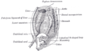 The primitive mesentery of a six weeks’ human embryo, half schematic.

References
- ^ a b c d Mitchell, Barry; Sharma, Ram (2009-01-01), Mitchell, Barry; Sharma, Ram (eds.), "Chapter 3 - The body cavities and the diaphragm", Embryology (Second Edition), Churchill Livingstone, pp. 15–18, doi:10.1016/b978-0-7020-3225-7.50006-3, ISBN 978-0-7020-3225-7, retrieved 2020-12-05
- ^ Moore, Keith L. (2003). The developing human : clinically oriented embryology. Persaud, T. V. N. (7th [ed.] ed.). Philadelphia, Pa.: Saunders. ISBN 978-0-8089-2265-0. OCLC 49526919.
- ^ Moore, N. Anthony. (2007). Gross and developmental anatomy. Roy, William A. (2nd ed.). St. Louis: Mosby. ISBN 978-0-323-04551-3. OCLC 70220003.
- ^ Carlson, Bruce M. (2004). Human embryology and developmental biology. Carlson, Bruce M. (3rd ed.). [St. Louis, Mo.]: Mosby. ISBN 0-323-01487-9. OCLC 60346934.
External links
- digest-016—Embryo Images at University of North Carolina
- mslimb-011—Embryo Images at University of North Carolina
- cardev-007—Embryo Images at University of North Carolina
- Fisher, Jason C; Bodenstein, Lawrence (December 2006). "Computer simulation analysis of normal and abnormal development of the mammalian diaphragm". Theoretical Biology and Medical Modelling. 3 (1): 9. doi:10.1186/1742-4682-3-9. PMC 1434728. PMID 16483386.