
| Short posterior ciliary arteries | |
|---|---|
 The arteries of the choroid and iris. The greater part of the sclera has been removed. | |
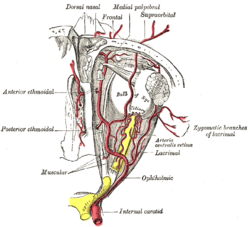 The ophthalmic artery and its branches | |
| Details | |
| Source | Ophthalmic artery |
| Vein | Vorticose veins |
| Supplies | Choroid (up to the equator of the eye) ciliary processes |
| Identifiers | |
| Latin | arteriae ciliares posteriores breves |
| TA98 | A12.2.06.031 |
| TA2 | 4479 |
| FMA | 70777 |
| Anatomical terminology | |
The short posterior ciliary arteries are a number of branches of the ophthalmic artery. They pass forward with the optic nerve to reach the eyeball, piercing the sclera around the entry of the optic nerve into the eyeball.
YouTube Encyclopedic
-
1/3Views:48 5771 468122 639
-
Eyeball | Blood Supply
-
Ciliary ganglion
-
VISUAL PATHWAY ANIMATED ANATOMY VIDEO LECTURE
Transcription
Anatomy
The number of short posterior ciliary arteries varies between individuals;[1] one or more short posterior ciliary arteries initially branch off the ophthalmic artery,[2] subsequently dividing to form up to 20 short posterior ciliary arteries.[2][3]
Origin
The short posterior ciliary arteries branch off the ophthalmic artery as it crosses the optic nerve medially.[1]
Course and relations
About 7 short posterior ciliary arteries accompany the optic nerve,[3] passing anterior-ward to reach the posterior part of[4] the eyeball, where they divide into 15-20 branches and pierce the sclera[3] around the entrance of the optic nerve.[4][2]
Distribution
The short posterior ciliary arteries contribute arterial supply to the choroid, ciliary processes,[3] optic disc, the outer retina, and Bruch's membrane.[1]
Some branches of the short posterior ciliary arteries supply the optic disc by means of an anastomotic ring - the circle of Zinn-Haller or circle of Zinn - which is associated with the fibrous extension of the ocular tendons (common tendinous ring (also annulus of Zinn)).[citation needed]
Additional images
-
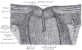 The terminal portion of the optic nerve and its entrance into the eyeball, in horizontal section

See also
References
- ^ a b c Gupta, Neha; Motlagh, Mahsaw; Singh, Gurdeep (2022), "Anatomy, Head and Neck, Eye Arteries", StatPearls, Treasure Island (FL): StatPearls Publishing, PMID 30725748, retrieved 2023-01-02
- ^ a b c Remington, Lee Ann (2012). "Orbital Blood Supply". Clinical Anatomy and Physiology of the Visual System. Elsevier. pp. 202–217. doi:10.1016/b978-1-4377-1926-0.10011-6. ISBN 978-1-4377-1926-0.
- ^ a b c d Standring, Susan (2020). Gray's Anatomy: The Anatomical Basis of Clinical Practice (42nd ed.). [New York]. p. 780. ISBN 978-0-7020-7707-4. OCLC 1201341621.
{{cite book}}: CS1 maint: location missing publisher (link) - ^ a b Gray, Henry (1918). Gray's Anatomy (20th ed.). p. 571.
This cardiovascular system article is a stub. You can help Wikipedia by expanding it. |