
| Radial collateral ligament | |
|---|---|
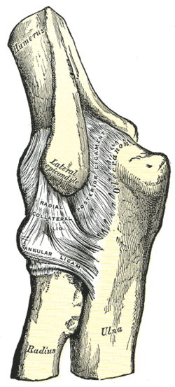 Left elbow joint, showing posterior and radial collateral ligaments. (Radial collateral ligament visible near center.) | |
| Details | |
| From | lateral epicondyle |
| To | annular ligament |
| Identifiers | |
| Latin | ligamentum collaterale radiale |
| TA98 | A03.5.09.006 |
| TA2 | 1777 |
| FMA | 38866 |
| Anatomical terminology | |

The radial collateral ligament (RCL), lateral collateral ligament (LCL), or external lateral ligament[Explain 1] is a ligament in the elbow on the side of the radius.
YouTube Encyclopedic
-
1/3Views:124 55726 0946 635
-
Elbow Joint - Human Anatomy | Kenhub
-
Ligaments of the Elbow
-
Fucntions of the Elbow Ligaments
Transcription
Hello everyone! This is Joao from Kenhub, and today, of course, I’m bringing to you another anatomy tutorial, this time, dedicated to another joint in your body, another tricky one to learn, and this is the elbow joint as you probably guess by looking at this image here. Now, I’m going to simplify or try to simplify this joint to you because it’s quite tricky to learn, and for that reason, the very first thing that you have to understand is that the elbow joint is happening between three bones, three main bones as you notice here on this image as well. Now, the first one is this large bone here known as the humerus, and then you have this one located laterally, and this is the radius. And the third bone is this one located medially. This is the ulna. So these are the three bones that comprise the elbow joint or that are in the elbow joint. Now, an important thing to know is that also the elbow joint is a synovial hinge joint, and it is a compound joint. What is a compound joint? That might be one of the questions that you have in your mind right now. And a compound joint is simply a joint that is comprised of other joints, other sub-joints—I use this term loosely—but other sub-joints. And the sub-joints, let’s say, that belong to the elbow joint or in the elbow joint are the humeroradial joint. As the name indicates, this is the joint occurring between the humerus, more specifically the capitulum of the humerus and the head of the radius. So this is the humeroradial joint. Now, the other one is, of course, the humeroulnar joint, and this is also happening, as the name indicates, with the trochlea of the humerus. This structure right here that I’m showing to you, and then of course, the trochlear notch of the ulna. Now, there is another joint that belongs—the third joint—that belongs to the elbow joint, and this is known as the proximal radioulnar joint, and this is a joint or an articulation that is occurring between the circumference or the articular circumference of the head of the radius, as you can see here, and then the radial notch of the ulna right here. So this is the proximal radioulnar joint. Keep in mind that I want to dedicate another tutorial, and I’m going to do that later, on this joint itself, the proximal radioulnar joint because you also find another one a little bit more distally known as the distal radioulnar joint, also occurring between the radius and the ulna. Now, knowing that, another point that I want to add here on the articular surfaces of the elbow joint of all these three joints that belong to the elbow joint is that they’re all covered or the articular surfaces are all covered, as you can see here on this image—this lighter or grayish color which is representing hyaline cartilage. So all the articular surfaces of the elbow joint are covered by hyaline cartilage. Now, we’re going to talk about the capsule of the elbow joint, and I’m going to show you it right now. So this is the capsule. And the capsule also has an outline and is attaching to several structures on each bone that we talked about. So as you see on this outline here above or proximally, the capsule is attaching to the margins or along the margins of the coronoid and also the radial fossae. And I can just show you by removing the capsule that these are the margins of—here, this is the radial fossa, and here is the margin of the coronoid fossa, the humerus. Now, if I put it back, you can also see that the capsule is attaching to both the lateral epicondyle and also the medial epicondyle of the humerus. But keep in mind that both epicondyles are exposed, as you can see here, so they’re not contained within this capsule that we’re talking about. Now, a little bit more distally or a little bit below here, you notice that the capsule is attached to the margin of the coronoid process here on the ulna. And it is attached on the radial side too on the lateral side to a ligament that we’re going to talk about and I’m showing you right now. So I just added this image of this ligament known as the annular ligament, and the capsule is definitely attaching to this ligament as well here on the lateral side. Now, we’re still talking about the capsule of the elbow joint, but this time, we’re looking at it on the posterior side or the back view of the elbow joint. And I just have here an outline in green of where the joint should be or the capsule of the joint should be. So as you notice here that on the proximal side or this upper view here, that it is attached to the margins of the olecranon fossa of the humerus. Now, if you go a little bit more distally, you’ll notice here that it’s also attached to other margins, but this time, of the olecranon process of the ulna. Also a little bit more distally and you notice here as I’m pointing it right now, there should be an attachment that will happen on the annular ligament that I just showed you before. So this is the posterior view of the capsule of the elbow joint. Now, as I mentioned before, I told you that the elbow joint is a synovial hinge joint, and synovial means that this joint will have space or there will be space between the articulating surfaces. And for that reason, you will find, then, what we’re going to talk about right now—the synovial membrane. So there is a synovial membrane and I’m showing you right now the outline of this synovial membrane that is clearly lining the capsule of the elbow joint as you can see here. So the synovial membrane is lining the capsule. And also, a couple of things that I’m going to add here that you’ll find on the synovial membrane is that it covers fatty pads in the coronoid, the radial, and also the olecranon fossae. And also, you can notice that it goes a little bit beyond or continuously below the synovial membrane of that other sub-joint that I talked about, the proximal radioulnar joint. Now, we’re going to talk about very important structures that help stabilize the elbow joint, and these are the ligaments, and there are three ligaments that we need to briefly go over and talk about on this tutorial. The first one I already discussed and mentioned a little bit about it. This was, then, the annular ligament. And as you can see here, the annular ligament is surrounding the head of the radius and attaches on the ulna on both sides. Now, the other two—there are two very important ligaments known as collateral ligaments. They’re very, very strong. They’re main stabilizers of the elbow joint. And the first one that I’m going to show you right now, this is known as the ulnar collateral ligament or you can also call it the medial collateral or the medial ligament. We’re going to discuss a little bit more in detail on the next slide. But for now, you can see here, highlighted in blue that this is the ulnar collateral ligament. Now, the other one is then going to be the radial collateral ligament here, highlighted in yellow, and this is another very strong ligament that we’re going to briefly discuss as well. Now, I wanted to talk a little bit more about the collateral ligaments, and let’s start, then, with the radial collateral ligament. This yellow structure here. And what I want to say is this is a triangular shaped ligament. You cannot see very well on this image here, but you can see enough for me to tell you that this is attaching to the tip, as you notice here, of the lateral epicondyle of the humerus then goes all the way, if you follow, this is all the way to the upper margin of this red ligament that we just talked about, the annular ligament. Now, a few words on the ulnar collateral ligament, this structure here highlighted in blue—as you notice here, the ulnar collateral ligament has a slightly triangular shape that you can notice here on this image, and it is comprised of three very, very strong bands. And in order for us to understand how they look, and how they are in comparison to one another, and how they’re attached, we’re going to use a very, very simple image here that, sometimes, we have to rely on whenever we’re studying anatomy. We have to simplify things and make it a little bit more clearer. So this image here is showing, as you can see, the triangular-shaped ulnar collateral ligament, and the three different bands are, of course, in different colors, in different blue colors. Now, here, you have the humerus, and of course, the ulna. Let me tell you that you’re having a medial view here, so you’re looking at it as if you were looking at this direction. So this image here on your left is the anterior view, and now, you’re looking at the collateral ligament, the ulnar collateral ligament on a medial side, medial view. And you would see, of course, this clear triangular shape that the ligament has. Now, the first band that you can notice here is located a little bit on the anterior side, and of course, you would call it, then, the anterior band. And as you can see, it’s attaching from the medial epicondyle to the medial margin of the coronoid process of the ulna. Now, you see another band here in a darker blue, and this band is then the posterior band, and the posterior band is attaching from also the medial epicondyle of the humerus right here and goes all the way to attach on this side, on the medial side of the olecranon of the ulna. Now, the third band is this one right here, another blue, and this is known as the transverse band. And as you can see, this transverse band is connecting, in a way, the two other bands that we talked about. So this transverse band is between the ulnar attachments of the two other bands that we talked about. Now, on the last part of this tutorial, I’m just going to briefly mention a couple of words on nerve supply of the elbow joint, and I can tell you that there will be four main nerves that also support or supply the arm and the forearm, and branches of these nerves will then supply the structures found on the elbow joint. Now, the first one that you need to know is definitely the median nerve. Also, branches of the ulnar nerve and the radial nerve will be supplying the elbow joint. And finally, the fourth nerve that will be supplying this joint is going to be the musculocutaneous nerve. All these four nerves have branches that will then supply the elbow joint. Now that you just completed this video tutorial, then it’s time for you to continue your learning experience by testing and also applying your knowledge. There are three ways you can do so here at Kenhub. The first one is by clicking on our “start training” button, the second one is by browsing through our related articles library, and the third one is by checking out our atlas. Now, good luck everyone, and I will see you next time. https://www.kenhub.com
Structure
The composition of the triangular ligamentous structure on the lateral side of the elbow varies widely between individuals[1] and can be considered either a single ligament,[2] in which case multiple distal attachments are generally mentioned and the annular ligament is described separately, or as several separate ligaments,[1] in which case parts of those ligaments are often described as indistinguishable from each other.
In the latter case, the ligaments are collectively referred to as the lateral collateral ligament complex (LCLC), consisting of four ligaments:[1]
- the radial collateral ligament [proper] (RCL), from the lateral epicondyle to the annular ligament deep to the common extensor tendon[1]
- the lateral ulnar collateral ligament (LUCL), from the lateral epicondyle[3] to the supinator crest on the ulna. Near the attachment on the humerus this ligament is normally indistinguishable from the RCL and can be considered the posterior portion of it.[1] Martin 1958 described the distal part of the LUCL as "a definite bundle which normally crosses the annular band and gains attachment to the supinator crest, frequently to a special tubercle on that crest" but didn't name it.[4]
- the annular ligament (AL), from the posterior to the anterior margins of radial notch on the ulna, encircles the head of radius and holds it against the radial notch of ulna.[5]
- the accessory lateral collateral ligament (ALCL), from the inferior margin of the annular ligament to the supinator crest.
Clinical significance
The radial collateral ligament may be involved in lateral epicondylitis.[6]
Additional images
-
 Elbow joint. Deep dissection. Anterior view.
 -
 Elbow joint. Deep dissection. Anterior view.
Explanations
- ^ As opposed to the "internal lateral ligament", better known as the medial or ulnar collateral ligament
References
- ^ a b c d e Carrino et al. 2001, Discussion, see also Figure 4
- ^ Palastanga & Soames 2012, Radial collateral ligament, p.133
- ^ "The Elbow Joint". TeachMeAnatomy. 2012-03-20. Retrieved 2017-07-24.
- ^ de Haan et al. 2011, Results
- ^ "Radio-Ulnar Joints". Earth's Lab. Retrieved 2017-11-18.
- ^ Jacobson, Jon A.; Chiavaras, Mary M.; Lawton, Jason Michael; Downie, Brian; Yablon, Corrie M.; Lawton, Jeffrey (2014). "Radial Collateral Ligament of the Elbow". Journal of Ultrasound in Medicine. 33 (6): 1041–1048. doi:10.7863/ultra.33.6.1041. hdl:2027.42/135312. ISSN 1550-9613.
Bibliography
- Carrino JA, Morrison WB, Zou KH, Steffen RT, Snearly WN, Murray PM (January 2001). "Lateral Ulnar Collateral Ligament of the Elbow: Optimization of Evaluation with Two-dimensional MR Imaging". Radiology. 218 (1): 118–25. doi:10.1148/radiology.218.1.r01ja52118. PMID 11152789.
- de Haan J, Schep NW, Eygendaal D, Kleinrensink GJ, Tuinebreijer WE, den Hartog D (2011). "Stability of the Elbow Joint: Relevant Anatomy and Clinical Implications of In Vitro Biomechanical Studies". The Open Orthopaedics Journal. 5: 168–76. doi:10.2174/1874325001105010168. PMC 3104563. PMID 21633722.
- Martin, BF (July 1958). "The annular ligament of the superior radio-ulnar joint". J. Anat. 92 (Pt3): 473–82. PMC 1245018. PMID 13563324.
- Palastanga, Nigel; Soames, Roger (2012). Anatomy and Human Movement: Structure and Function (6th ed.). Elsevier. ISBN 9780702040535.