
| Quadratus plantae muscle | |
|---|---|
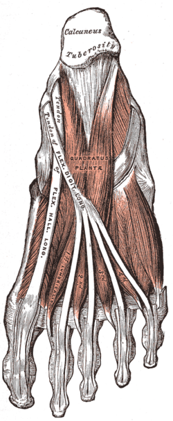 Muscles of the sole of the foot. Second layer. (Quadratus plantae visible at center.) | |
| Details | |
| Origin | Calcaneus, Long plantar ligament |
| Insertion | Tendons of Flexor digitorum longus |
| Nerve | Lateral plantar nerve (S1,2) |
| Actions | Assists flexor digitorum longus in flexion of DIP joints |
| Identifiers | |
| Latin | musculus quadratus plantae |
| TA98 | A04.7.02.068 |
| TA2 | 2684 |
| FMA | 37452 |
| Anatomical terms of muscle | |
The quadratus plantae (flexor accessorius) is separated from the muscles of the first layer by the lateral plantar vessels and nerve. It acts to aid in flexing the 2nd to 5th toes (offsetting the oblique pull of the flexor digitorum longus) and is one of the few muscles in the foot with no homolog in the hand.
YouTube Encyclopedic
-
1/3Views:225 88731 743655
-
Nerves Of The Leg & Foot - Everything You Need To Know - Dr. Nabil Ebraheim
-
Foot Muscles Anatomy part 1/2
-
BYUH Anatomy Lab- Lower leg Muscles Peroneous Brevis to Quadratratus Plantae
Transcription
Origin and insertion
It arises by two heads, which are separated from each other by the long plantar ligament: the medial or larger head is muscular, and is attached to the medial concave surface of the calcaneus, below the groove which lodges the tendon of the flexor hallucis longus; the lateral head, flat and tendinous, arises from the lateral border of the inferior surface of the calcaneus, in front of the lateral process of its tuberosity, and from the long plantar ligament.
The two portions join at an acute angle, and end in a flattened band which is inserted into the lateral margin and upper and under surfaces of the tendon of the flexor digitorum longus, forming a kind of groove, in which the tendon is lodged. It usually sends slips to those tendons of the flexor digitorum longus which pass to the second, third, and fourth toes.
Variations
Lateral head often wanting; entire muscle absent. Variation in the number of digital tendons to which fibers can be traced. Most frequent offsets are sent to the second, third and fourth toes; in many cases to the fifth as well; occasionally to two toes only.
Additional images
-
 Bones of the right foot. Plantar surface.
 -
 Coronal section through right talocrural and talocalcaneal joints
 -
 The plantar arteries. Deep view. (Quadratus plantae visible at center.)
 -
 The quadratus plantae aids in flexion of the toes


References
![]() This article incorporates text in the public domain from page 493 of the 20th edition of Gray's Anatomy (1918)
This article incorporates text in the public domain from page 493 of the 20th edition of Gray's Anatomy (1918)
External links
This muscle article is a stub. You can help Wikipedia by expanding it. |