
| Oblique popliteal ligament | |
|---|---|
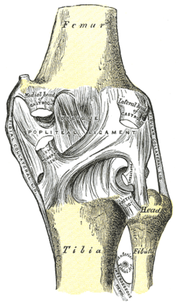 Right knee-joint. Posterior view. (Oblique popliteal ligament visible at center.) | |
| Details | |
| From | lateral epicondyle of the femur, lateral condyle of femur |
| To | medial condyle of tibia |
| Identifiers | |
| Latin | ligamentum popliteum obliquum |
| TA98 | A03.6.08.013 |
| TA2 | 1899 |
| FMA | 44582 |
| Anatomical terminology | |
The oblique popliteal ligament (posterior ligament) is a broad, flat, fibrous ligament on the posterior knee.[1] It is an extension of the tendon of the semimembranosus muscle.[1][2] It attaches onto the intercondylar fossa and lateral condyle of the femur.[2] It reinforces the posterior central portion of the knee joint capsule.[3]
YouTube Encyclopedic
-
1/5Views:49 400306 6661 0659 22774 588
-
Knee Joint - 4 | Arcuate Popliteal Ligament | Oblique Popliteal Ligament
-
LIGAMENTS OF THE KNEE
-
Anatomy of posterior oblique ligament of knee joint (English)
-
Knee Anatomy Tutorial (Ligaments)
-
Popliteus Tendon Evaluation [Back of the knee pain]
Transcription
Anatomy
The oblique popliteal ligament is formed as a lateral expansion of the tendon of the semimembranosus muscle[2] and represents one of the muscle's five insertions.[citation needed] The ligament blends with the posterior portion of the knee joint capsule.[2] It exhibits a large opening through which nerves and vessels pass.[3]
Attachments
The ligament extends superolaterally from the semimembranosus tendon to attach onto the intercondylar fossa and lateral condyle of the femur.[2][3]
Relations
The oblique popliteal ligament forms part of the floor of the popliteal fossa;[citation needed] the popliteal artery lies upon the ligament.[2] The ligament is pierced by posterior division of the obturator nerve, as well as the middle genicular nerve, the middle genicular artery, and the middle genicular vein.[citation needed]
Clinical significance
The oblique popliteal ligament may be damaged, causing a valgus deformity. Surgical repair of the ligament often leads to better outcomes than conservative management.[4]
The oblique popliteal ligament may be cut during arthroscopic meniscus repair surgery.[5]
Additional images
-
 Sagittal section of right knee-joint.

References
![]() This article incorporates text in the public domain from page 340 of the 20th edition of Gray's Anatomy (1918)
This article incorporates text in the public domain from page 340 of the 20th edition of Gray's Anatomy (1918)
- ^ a b Mehta, V; Dawani, P; Goel, P (September 2022). "Morphologic and Morphometric Evaluation of Oblique Popliteal Ligament - A Clinico-Anatomical Study". Maedica. 17 (3): 641–646. doi:10.26574/maedica.2022.17.3.641 (inactive 31 January 2024). PMC 9720660. PMID 36540577.
{{cite journal}}: CS1 maint: DOI inactive as of January 2024 (link) - ^ a b c d e f Chummy S. Sinnatamby (2011). Last's anatomy: regional and applied (12th ed.). Edinburgh. p. 138. ISBN 978-0-7020-4839-5. OCLC 764565702.
{{cite book}}: CS1 maint: location missing publisher (link) - ^ a b c Palastanga, Nigel; Soames, Roger (2012). Anatomy and Human Movement: Structure and Function. Physiotherapy Essentials (6th ed.). Edinburgh: Churchill Livingstone/Elsevier. p. 307. ISBN 978-0-7020-3553-1.
- ^ Berkson, Eric M.; Nolan, David; Fleming, Kristina; Spang, Robert; Wong, Jeff; Asnis, Peter; Kawadler, Jaeson (2016-01-01), Magee, David J.; Zachazewski, James E.; Quillen, William S.; Manske, Robert C. (eds.), "Knee", Pathology and Intervention in Musculoskeletal Rehabilitation (Second Edition), W.B. Saunders, pp. 713–773, doi:10.1016/b978-0-323-31072-7.00020-8, ISBN 978-0-323-31072-7, retrieved 2021-03-02
- ^ Nawab, Akbar; Hester, Peter W.; Caborn, David N. M. (2004-01-01), Miller, Mark D.; Cole, Brian J.; Cohen, Steven B.; Makda, Junaid A. (eds.), "Arthroscopic Meniscus Repair", Textbook of Arthoscopy, Philadelphia: W.B. Saunders, pp. 517–537, doi:10.1016/b978-0-7216-0013-0.50054-5, ISBN 978-0-7216-0013-0, retrieved 2021-03-02
External links
- Oblique popliteal ligament at the Duke University Health System's Orthopedics program
- lljoints at The Anatomy Lesson by Wesley Norman (Georgetown University) (postkneejointsuperfic)
- Anatomy photo:17:02-0400 at the SUNY Downstate Medical Center - "Major Joints of the Lower Extremity: Knee Joint"
This ligament-related article is a stub. You can help Wikipedia by expanding it. |