Ege Üniversitesi Doğa Tarihi Müzesi | |
| Established | 1973 |
|---|---|
| Coordinates | 38°27′31″N 27°13′53″E / 38.45861°N 27.23139°E |
| Type | Natural History |
| Collection size | about 4000[1] |
| Owner | Ege University |
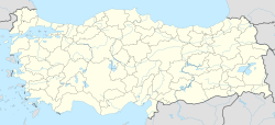
Natural History Museum of Ege University (Turkish: Ege Universitesi Doğa Tarihi Müzesi) is a university museum in İzmir, Turkey. The museum is in the campus of the Ege University next to Faculty of Science at 38°27′31″N 27°13′53″E / 38.45861°N 27.23139°E. The museum was founded in 1973. Between 1973 and 1991 it was an institute of the faculty. After 1991 it became a subunit of the rectorship.
YouTube Encyclopedic
-
1/3Views:2 765 2291 466483
-
Flow through the heart | Circulatory system physiology | NCLEX-RN | Khan Academy
-
2015 CSULB Commencement - Natural Sciences & Mathematics
-
Utveksling til University of Hong Kong
Transcription
So what you're looking at is one of the most amazing organs in your body. This is the human heart. And it's shown with all the vessels on it. And you can see the vessels coming into it and out of it. But the heart, at its core, is a pump. And this pump is why we call it the hardest working organ in our body. Because it starts pumping blood from the point where you're a little fetus, maybe about eight weeks old, all the way until the point where you die. And so this organ, I think, would be really cool to look at in a little bit more detail. But it's hard to do that looking just at the outside. So what I did is I actually drew what it might look like on the inside. So let me actually just show you that now. And we'll follow the path of blood through the heart using this diagram. Let me start with a little picture in the corner. So let's say we have a person here. And this is their face, and this is their neck. I'm going to draw their arms. And they have, in the middle of their chest, their heart. And so the whole goal is to make sure that blood from all parts of their body, including their legs, can make its way back to the heart, first of all, and then get pumped back out to the body. So blood is going to come up from this arm, let's say, and dump into there. And the same on this side. And it's going to come from their head. And all three sources, the two arms and the head, are going to come together into one big vein. And that's going to be dumping into the top of the heart. And then separately, you've got veins from the legs meeting up with veins from the belly, coming into another opening into the heart. So that's how the blood gets back to the heart. And any time I mention the word vein, I just want you to make sure you think of blood going towards the heart. Now if blood is going towards the heart, then after the blood is pumped by the heart, it's going to have to go out to the heart. It's going to have to go away from the heart. So that's the aorta. And the aorta actually has a little arch, like that. We call it the aortic arch. And it sends off one vessel to the arm, one vessel up this way, a vessel over this way. And then this arch kind of goes down, down, down and splits like that. So this is kind of a simplified version of it. But you can see how there are definitely some parallels between how the veins and the arteries are set up. And arteries, anytime I mention the word artery, I want you to think of blood going away from the heart. And an easy way to remember that is that they both start with the letter A. So going to our big diagram now. We can see that blood coming in this way and blood coming in this way is ending up at the same spot. It's going to end up at the-- actually, maybe I'll draw it here-- is ending up at the right atrium. That's just the name of the chamber that the blood ends up in. And it came into the right atrium from a giant vessel up top called the superior vena cava. And this is a vein, of course, because it's bringing blood towards the heart. And down here, the inferior vena cava. So these are the two directions that blood is going to be flowing. And once blood is in the right atrium, it's going to head down into the right ventricle. So this is the right ventricle, down here. This is the second chamber of the heart. And it gets there by passing through a valve. And this valve, and all valves in the heart, are basically there to keep blood moving in the right direction. So it doesn't go in the backwards direction. So this valve is called the tricuspid valve. And it's called that because it's basically got three little flaps. That's why they call it tri. And I know you can only see two in my drawing, and that's just because my drawing is not perfect. And it's hard to show a flap coming out at you, but you can imagine it. So blood goes into the right ventricle. And where does it go next? Well after that, it's going to go this way. It's going to go into this vessel, and it's going to split. But before it goes there, it has to pass through another valve. So this is a valve, right here, called the pulmonary valve. And it gives you a clue as to where things are going to go next. Right? Because the word pulmonary means lungs. And so, if this is my lung, on this side, this is my left lung. And this is my right lung, on this side. Then these vessels-- and I'll let you try to guess what they would be called-- these vessels. This would be my-- I want to make sure I get my right and left straight. This is my left pulmonary artery. And I hesitated there just to make sure you got that because it's taking blood away from the heart. And this is my right pulmonary artery. So this is my right and left pulmonary artery. And so blood goes, now, into my lungs. These are the lungs that are kind of nestled into my thorax, where my heart is sitting. It goes into my lungs. And remember, this blood is blue. Why is it blue? Well, it's blue because it doesn't have very much oxygen. And so one thing that I need to pick up is oxygen. And so that's one thing that the lungs are going to help me pick up. And I'm going to write O2 for oxygen. And it's also blue. And that reminds us that it's full of carbon dioxide. It's full of waste because it's coming from the body. And the body's made a lot of carbon dioxide that it's trying to get rid of. So in the lungs, you get rid of your carbon dioxide and you pick up oxygen. So that's why I switch, at this point, from a blue-colored vessel to a red-colored vessel. So now blood comes back in this way and this way and dumps into this chamber. So what is that? This is our left atrium. So just like our right atrium, we have one on the left. And it goes down into-- and you can probably guess what this one is called-- it's our left ventricle. So just like before, where it went from the right atrium to the right ventricle, now we're going from the left atrium to the left ventricle. And it passes through a valve here. So this valve is called the mitral valve. And its job is, of course, to make sure that blood does not go from the left ventricle back to the left atrium by accident. It wants to make sure that there's forward flow. And then the final valve-- I have to find a nice spot to write it, maybe right here. This final valve that it passes through is called the aortic valve. And the aortic valve is going to be what divides the left ventricle from this giant vessel that we talked about earlier. And this is, of course, the aorta. This is my aorta. So now blood is going to go through the aorta to the rest of the body. So you can see how blood now flows from the body into the four chambers. First into the right atrium-- this is chamber number one. And then it goes into the right ventricle. This is chamber number two. It goes to the lungs and then back out to the left atrium. So this is chamber number three. And then the left ventricle. And this happens every moment of every day. Every time you hear your heart beating, this process is going on.
Exhibited items
There are 4000 items in the exhibition. There are 6 halls in 2,500 square metres (27,000 sq ft) area.[1]
- 1. Paleontology (1168 items)
- 2. Rocks and minerals (811 items)
- 3. Birds (168 items)
- 4. Entrance (Turkish fauna) (937 items)
- 5. General zoology (766 items)
- 6. Osteology and evolution (81 items)


