
| Medial septal nucleus | |
|---|---|
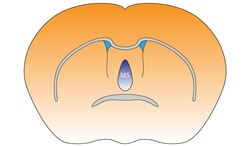 Medial septal nucleus of the mouse brain | |
| Details | |
| Identifiers | |
| Latin | nucleus septalis medialis |
| NeuroNames | 262 |
| NeuroLex ID | birnlex_1668 |
| TA98 | A14.1.09.269 A14.1.09.446 |
| FMA | 61879 |
| Anatomical terms of neuroanatomy | |
The medial septal nucleus (MS) is one of the septal nuclei. Neurons in this nucleus give rise to the bulk of efferents from the septal nuclei. A major projection from the medial septal nucleus terminates in the hippocampal formation.[1]
It plays a role in the generation of theta waves in the hippocampus.[2] Specifically, the GABAergic cells of the medial septum that act as theta pacemakers target dentate gyrus, CA3, and CA1 interneurons. Pacemaking MS interneurons express hyperpolarization-activated cyclic nucleotide-gated (HCN) channels which likely, at least partially, mediate their pacemaker properties.[3]
It is composed of GABAergic cells, glutamatergic cells, and cholinergic cells. Each cell-type carries out different functions. In addition to the theta wave generation, it has recently been discovered that medial septum also serves as an important node for sensory valence processing. For example, Vglut2 neurons in medial septum respond strongly to noxious sensory stimulation.
YouTube Encyclopedic
-
1/3Views:1 601 01617 96347 168
-
Cardiac Conduction System and Understanding ECG, Animation.
-
Fornix of the brain: structure and function - Human Anatomy | Kenhub
-
Brain Anatomy - Brain Fornix and Ventricle Anatomy
Transcription
The cardiac conduction system consists of the following components: - The sinoatrial node, or SA node, located in the right atrium near the entrance of the superior vena cava. This is the natural pacemaker of the heart. It initiates all heartbeat and determines heart rate. Electrical impulses from the SA node spread throughout both atria and stimulate them to contract. - The atrioventricular node, or AV node, located on the other side of the right atrium, near the AV valve. The AV node serves as electrical gateway to the ventricles. It delays the passage of electrical impulses to the ventricles. This delay is to ensure that the atria have ejected all the blood into the ventricles before the ventricles contract. - The AV node receives signals from the SA node and passes them onto the atrioventricular bundle - AV bundle or bundle of His. - This bundle is then divided into right and left bundle branches which conduct the impulses toward the apex of the heart. The signals are then passed onto Purkinje fibers, turning upward and spreading throughout the ventricular myocardium. Electrical activities of the heart can be recorded in the form of electrocardiogram, ECG or EKG. An ECG is a composite recording of all the action potentials produced by the nodes and the cells of the myocardium. Each wave or segment of the ECG corresponds to a certain event of the cardiac electrical cycle. When the atria are full of blood, the SA node fires, electrical signals spread throughout the atria and cause them to depolarize. This is represented by the P wave on the ECG. Atrial contraction , or atrial systole starts about 100 ms after the P wave begins. The P-Q segment represents the time the signals travel from the SA node to the AV node. The QRS complex marks the firing of the AV node and represents ventricular depolarization: - Q wave corresponds to depolarization of the interventricular septum. - R wave is produced by depolarization of the main mass of the ventricles. - S wave represents the last phase of ventricular depolarization at the base of the heart. - Atrial repolarization also occurs during this time but the signal is obscured by the large QRS complex. The S-T segment reflects the plateau in the myocardial action potential. This is when the ventricles contract and pump blood. The T wave represents ventricular repolarization immediately before ventricular relaxation, or ventricular diastole. The cycle repeats itself with every heartbeat.
References
- ^ Brodal, A (1981). Neurological Anatomy. New York, N.Y.: Oxford University Press. Retrieved 18 Jun 2011.
- ^ O'Keefe, John; Andersen, Per; Morris, Richard; David Amaral; Tim Bliss (2007). The hippocampus book. Oxford [Oxfordshire]: Oxford University Press. p. 480. ISBN 978-0-19-510027-3.
- ^ Colgin, Laura Lee (April 2016). "Rhythms of the hippocampal network". Nature Reviews. Neuroscience. 17 (4): 239–249. doi:10.1038/nrn.2016.21. ISSN 1471-003X. PMC 4890574. PMID 26961163.
This neuroanatomy article is a stub. You can help Wikipedia by expanding it. |