
| Lobules of testis | |
|---|---|
 A diagram of the major components of an adult human testicle, including the following numbered items: 1. Tunica albuginea, 2. Septula testis, 3. Lobulus testis, 4. Mediastinum testis, 5. Tubuli seminiferi contorti, 6. Tubuli seminiferi recti, 7. Rete testis, 8. Ductuli efferentes testis, 9a. Head of epididymis, 9b. Body of epididymis, 9c. Tail of epididymis, 10. Vas deferens, 11a. Tunica vaginalis (parietal lamina), 11b. Tunica vaginalis (visceral lamina), and 12. Cavity of tunica vaginalis. | |
 1: Head or upper pole of testis, 2: Tunica albuginea, 3: Testicular septa, 4: Anterior margin (free margin), 5: Lateral surface, 6: Tail or lower pole of testis, 7: Testicular lobules, 8: Parenchyma of testis, 9: Efferent ductules, 10: Mediastinum testis, 11: Posterior margin | |
| Details | |
| Identifiers | |
| Latin | lobuli testis |
| Anatomical terminology | |
The lobules of testis are of partitions of the testis formed by septa of testis. The lobules of testis contain the tightly coiled seminiferous tubule.[1] There are some hundreds of lobules in a testicle.[2][3]
YouTube Encyclopedic
-
1/3Views:100 109400 7701 656
-
Testis and epididymis: structure and functions (preview) - Human Anatomy | Kenhub
-
Testis and Epididymis - Male Reproductive Anatomy Part 1
-
How many seminiferous tubules are present in each testicular lobule ?
Transcription
Anatomy
They differ in size according to their position, those in the middle of the gland being larger and longer.
The lobules are conical in shape, the base being directed toward the circumference of the organ, the apex toward the mediastinum testis.
Each lobule is contained in one of the intervals between the fibrous septa which extend between the mediastinum testis and the tunica albuginea, and consists of from one to three, or more, minute convoluted tubes, the seminiferous tubules (tubuli seminiferi).
Each tubule extends from the base of the lobule where the tubule ends blindly towards the apex of the lobule.[4][verification needed][better source needed]
Additional images
-
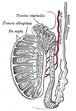 Vertical section of the testis, to show the arrangement of the ducts

References
![]() This article incorporates text in the public domain from the 20th edition of Gray's Anatomy (1918)
This article incorporates text in the public domain from the 20th edition of Gray's Anatomy (1918)
- ^ Martini, Frederic; Tallitsch, Robert B.; Nath, Judi L. (2017). Human Anatomy (9th ed.). Pearson. p. 711. ISBN 9780134320762.
- ^ Countouris, N; Holstein, AF (Nov–Dec 1985). "[How many testicular lobules does a human testicle contain? Reexamination of an old problem]". Andrologia (in German). 17 (6): 525–31. doi:10.1111/j.1439-0272.1985.tb01707.x. PMID 4083540. S2CID 86699538.
- ^ Basu, SC. (2011). Male Reproductive Dysfunction. Jaypee Brothers Publishers. p. 16. ISBN 978-93-5025-703-6.
- ^ DocCheck Medical Services GmbH. "Cavum vaginale". DocCheck Flexikon.
External links
- Anatomy photo:36:st-1401 at the SUNY Downstate Medical Center - "Inguinal Region, Scrotum and Testes: Testis"
≠