
| Lesser occipital nerve | |
|---|---|
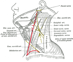 Side of neck, showing chief surface markings. (Lesser occip. nerve labeled at center right.) | |
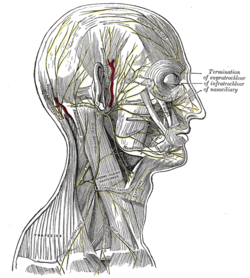 The nerves of the scalp, face, and side of neck. (Smaller occipital visible below and to the left of the ear.) | |
| Details | |
| From | cervical plexus (C2) |
| Innervates | Cutaneous innervation of the posterior aspect of the auricle and mastoid region |
| Identifiers | |
| Latin | nervus occipitalis minor |
| TA98 | A14.2.02.017 |
| TA2 | 6384 |
| FMA | 6871 |
| Anatomical terms of neuroanatomy | |
The lesser occipital nerve (or small occipital nerve[1]) is a cutaneous spinal nerve of the cervical plexus.[2] It arises from second cervical (spinal) nerve (C2) (along with the greater occipital nerve). It innervates the skin of the back of the upper neck and of the scalp posterior to the ear.
YouTube Encyclopedic
-
1/3Views:60 31414 98949 126
-
Occipital Neuralgia/Splenius Irritation
-
Tinel's Sign - Greater Occipital Nerve
-
Scalp Blocks
Transcription
Structure
Origin
It arises from the (lateral branch of the ventral ramus[3][4]) of cervical spinal nerve C2;[3][4][2] it (sources differ) receives[1] or may also receive fibres from cervical spinal nerve C3.[3][4] It originates between the atlas, and axis.[4]
The lesser occipital nerve is one of the four cutaneous branches of the cervical plexus.[5]
Course and relations
It curves around the accessory nerve (CN XI)[4][3][6] to come to course anterior to it.[3] It then[3] curves around[4] and ascends along the posterior border of the sternocleidomastoid muscle;[3][4][6] rarely, it may pierce the muscle.[4] Near the cranium, it perforates the deep cervical fascia. It is continues upwards along the scalp posterior to the auricle.[3][4] It divides into medial and lateral segments between the inion, and intermastoid line.[1]
Branches
It has an auricular, a mastoid, and an occipital branch.[1]
Its auricular branch supplies the skin of the upper and back part of the auricula, communicating with the mastoid branch of the great auricular. This branch is occasionally derived from the greater occipital nerve.[citation needed]
Distribution
The nerve provides sensory innervation to the upper part of the back of the neck and adjacent[6] scalp posterior to the auricle;[6][1] it may also contribute to the sensory innervation of the auricle itself.[6]
Communications
It communicates with the greater occipital nerve,[4][1] great auricular nerve, and the auricular branch of the facial nerve.[4]
Variation
Rarely, the lesser occipital nerve may be duplicated or triplicated.[4]
Clinical significance
Problems with the lesser occipital nerve cause occipital neuralgia. Nerve block is difficult due to variation in the course of the nerve.[4]
Additional images
-
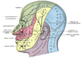
Dermatome distribution of the trigeminal nerve

References
- ^ a b c d e f Yu, Megan; Wang, Shu-Min (2023), "Anatomy, Head and Neck, Occipital Nerves", StatPearls, Treasure Island (FL): StatPearls Publishing, PMID 31194370, retrieved 2023-06-22
- ^ a b Sinnatamby, Chummy S. (2011). Last's Anatomy (12th ed.). Elsevier Australia. p. 334. ISBN 978-0-7295-3752-0.
- ^ a b c d e f g h Standring, Susan (2020). Gray's Anatomy: The Anatomical Basis of Clinical Practice (42nd ed.). [New York]. ISBN 978-0-7020-7707-4. OCLC 1201341621.
{{cite book}}: CS1 maint: location missing publisher (link) - ^ a b c d e f g h i j k l m Cesmebasi, Alper (2015). "31 - Anatomy of the Cervical Plexus and Its Branches". Nerves and Nerve Injuries. Vol. 1: History, Embryology, Anatomy, Imaging, and Diagnostics. Academic Press. pp. 441–449. doi:10.1016/B978-0-12-410390-0.00032-9. ISBN 978-0-12-410390-0.
- ^ Rea, Paul (2016). "3 - Neck". Essential Clinically Applied Anatomy of the Peripheral Nervous System in the Head and Neck. Academic Press. pp. 131–183. doi:10.1016/B978-0-12-803633-4.00003-X. ISBN 978-0-12-803633-4.
- ^ a b c d e Sinnatamby, Chummy S. (2011). Last's Anatomy (12th ed.). Elsevier Australia. p. 334. ISBN 978-0-7295-3752-0.
External links
- Anatomy figure: 25:03-01 at Human Anatomy Online, SUNY Downstate Medical Center
- http://www.dartmouth.edu/~humananatomy/figures/chapter_47/47-2.HTM
- http://www.dartmouth.edu/~humananatomy/figures/chapter_47/47-6.HTM