
| Internal pudendal veins | |
|---|---|
 | |
| Details | |
| Drains to | Internal iliac vein |
| Artery | Internal pudendal artery |
| Identifiers | |
| Latin | vena pudenda interna |
| TA98 | A12.3.10.019 |
| TA2 | 5032 |
| FMA | 18917 |
| Anatomical terminology | |
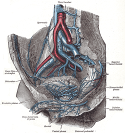
The internal pudendal veins (internal pudic veins) are a set of veins in the pelvis. They are the venae comitantes of the internal pudendal artery. Internal pudendal veins are enclosed by pudendal canal, with internal pudendal artery and pudendal nerve.
They begin in the deep veins of the vulva and of the penis and scrotum, issuing from the bulb of the vestibule and the bulb of the penis, respectively. They accompany the internal pudendal artery, and unite to form a single vessel, which ends in the internal iliac vein.
They receive the veins from the urethral bulb, the perineal and inferior hemorrhoidal veins.
The deep dorsal vein of the penis communicates with the internal pudendal veins, but ends mainly in the pudendal plexus.
YouTube Encyclopedic
-
1/3Views:12 613488 24867 440
-
Iliac Veins (Topography, Visceral and Parietal Tributaries) - Anatomy
-
Internal Iliac Artery
-
How to remember the Internal Iliac Artery Branches (The 2+4+4 rule) | Anatomy
Transcription
References
![]() This article incorporates text in the public domain from page 674 of the 20th edition of Gray's Anatomy (1918)
This article incorporates text in the public domain from page 674 of the 20th edition of Gray's Anatomy (1918)
External links
- Anatomy photo:13:06-0103 at the SUNY Downstate Medical Center - "Gluteal Region: Pudendal Nerve and Internal Pudendal Vessels"
- Anatomy photo:41:07-0105 at the SUNY Downstate Medical Center - "The Female Perineum: The Sacrotuberous and the Sacrospinous Ligaments"
This cardiovascular system article is a stub. You can help Wikipedia by expanding it. |