
| Golgi tendon organ | |
|---|---|
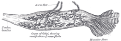 Labeled diagram of Golgi tendon organ from the human Achilles tendon. | |
| Details | |
| System | Musculoskeletal system |
| Location | Skeletal muscle |
| Identifiers | |
| Latin | organum sensorium tendinis |
| TH | H3.03.00.0.00024 |
| Anatomical terms of microanatomy | |
The Golgi tendon organ (GTO) (also called Golgi organ, tendon organ, neurotendinous organ or neurotendinous spindle) is a proprioceptor – a type of sensory receptor that senses changes in muscle tension. It lies at the interface between a muscle and its tendon known as the musculotendinous junction also known as the myotendinous junction.[1] It provides the sensory component of the Golgi tendon reflex.
The Golgi tendon organ is one of several eponymous terms named after the Italian physician Camillo Golgi.
YouTube Encyclopedic
-
1/5Views:163 93718 07368 82354 30061 729
-
Neurology | Spinal Cord: Golgi Tendon Organ Reflex (GTO)
-
Two Minutes of Anatomy: Golgi Tendon Organ
-
Reciprocal vs. Autogenic Inhibition Explained | Golgi Tendon Organ and Muscle Spindle Physiology
-
Muscle Sensors (Part II) - The Golgi Tendon Organ
-
Function of Golgi Tendon Organs [GTOs] in Movement & Exercise
Transcription
Structure
The body of the Golgi tendon organ is made up of braided strands of collagen (intrafusal fasciculi) that are less compact than elsewhere in the tendon and are encapsulated.[2] The capsule is connected in series (along a single path) with a group of muscle fibers (10-20 fibers[3]) at one end, and merge into the tendon proper at the other. Each capsule is about 1 mm long, has a diameter of about 0.1 mm, and is perforated by one or more afferent type Ib sensory nerve fibers (Aɑ fiber), which are large (12-20 μm) myelinated axons that can conduct nerve impulses very rapidly. Inside the capsule, the afferent fibers lose their medullary sheaths, branch, intertwine with the collagen fibers, and terminate as flattened leaf-like endings between the collagen strands (see figure).[4][5]
Function

When the muscle generates force, the sensory terminals are compressed. This stretching deforms the terminals of the Ib afferent axon, opening stretch-sensitive cation channels. As a result, the Ib axon is depolarized and fires nerve impulses that are propagated to the spinal cord. The action potential frequency signals the force being developed by 10-20 extrafusal muscle fibers in the muscle. Average level of activity in a tendon organ population is representative of the whole muscle force.[4][7]
The Ib sensory feedback generates stretch reflexes and supraspinal responses which control muscle contraction. Ib afferents synapse with interneurons in the spinal cord that also project to the brain cerebellum and cerebral cortex. The Golgi tendon reflex assists in regulating muscle contraction force. It is associated with the Ib. Tendon organs signal muscle force through the entire physiological range, not only at high strain.[7][8]
During locomotion, Ib input excites rather than inhibits motoneurons of the receptor-bearing muscles, and it affects the timing of the transitions between the stance and swing phases of locomotion.[9] The switch to autogenic excitation is a form of positive feedback.[10]
The ascending or afferent pathways to the cerebellum are the dorsal and ventral spinocerebellar tracts. They are involved in the cerebellar regulation of movement.[citation needed]
History
Until 1967 it was believed that Golgi tendon organs had a high threshold, only becoming active at high muscle forces. Consequently, it was thought that tendon organ input caused "weightlifting failure" through the clasp-knife reflex, which protected the muscle and tendons from excessive force. [citation needed] However, the underlying premise was shown to be incorrect by James Houk and Elwood Henneman in 1967.[11]
See also
Footnotes
Sources
![]() This article incorporates text in the public domain from page 1061 of the 20th edition of Gray's Anatomy (1918)
This article incorporates text in the public domain from page 1061 of the 20th edition of Gray's Anatomy (1918)
- ^ MacIntosh, Brian R. (2006). Skeletal muscle : form and function (2nd ed.). Champaign, IL: Human Kinetics. pp. 48–49. ISBN 0736045171.
- ^ Mancall, Elliott L; Brock, David G, eds. (2011). "Chapter 2 - Overview of the Microstructure of the Nervous System". Gray's Clinical Neuroanatomy: The Anatomic Basis for Clinical Neuroscience. Elsevier Saunders. p. 29. ISBN 978-1-4160-4705-6.
- ^ a b Purves et al (2018), Mechanoreceptors Specialized for Proprioception, pp. 201-202
- ^ a b Pearson & Gordon (2013), 35-3 Golgi Tendon Organs, p. 800
- ^ Saladin (2018), The Tendon Reflex, p. 498-499
- ^ Barrett, Kim E; Boitano, Scott; Barman, Susan M; Brooks, Heddwen L (2010). "Chapter 9 - Reflexes". Ganong's Review of Medical Physiology (23rd ed.). McGraw-Hill. INVERSE STRETCH REFLEX, pp. 162-163. ISBN 978-0-07-160567-0.
- ^ a b Prochazka, A.; Gorassini, M. (1998). "Ensemble firing of muscle afferents recorded during normal locomotion in cats". Journal of Physiology. 507 (1): 293–304. doi:10.1111/j.1469-7793.1998.293bu.x. PMC 2230769. PMID 9490855.
- ^ Stephens, J. A.; Reinking, R. M.; Stuart, D. G. (1975). "Tendon organs of cat medial gastrocnemius: responses to active and passive forces as a function of muscle length". Journal of Neurophysiology. 38 (5): 1217–1231. doi:10.1152/jn.1975.38.5.1217. PMID 1177014.
- ^ Conway, B. A.; Hultborn, H.; Kiehn, O. (1987). "Proprioceptive input resets central locomotor rhythm in the spinal cat". Experimental Brain Research. 68 (3): 643–656. doi:10.1007/BF00249807. PMID 3691733. S2CID 22961186.
- ^ Prochazka, A.; Gillard, D.; Bennett, D. J. (1997). "Positive Force Feedback Control of Muscles". J Neurophysiol. 77 (6): 3226–3236. doi:10.1152/jn.1997.77.6.3226. PMID 9212270.
- ^ Houk, J.; Henneman, E. (1967). "Responses of Golgi tendon organs to active contractions of the soleus muscle of the cat". Journal of Neurophysiology. 30 (3): 466–481. doi:10.1152/jn.1967.30.3.466. PMID 6037588.
Other sources
- Saladin, KS (2018). "Chapter 13 - The Spinal Cord, Spinal Nerves, and Somatic Reflexes". Anatomy and Physiology: The Unity of Form and Function (8th ed.). New York: McGraw-Hill. ISBN 978-1-259-27772-6.
- Purves, Dale; Augustine, George J; Fitzpatrick, David; Hall, William C; Lamantia, Anthony Samuel; Mooney, Richard D; Platt, Michael L; White, Leonard E, eds. (2018). "Chapter 9 - The Somatosensory System: Touch and Proprioception". Neuroscience (6th ed.). Sinauer Associates. ISBN 9781605353807.
- Pearson, Keir G; Gordon, James E (2013). "35 - Spinal Reflexes". In Kandel, Eric R; Schwartz, James H; Jessell, Thomas M; Siegelbaum, Steven A; Hudspeth, AJ (eds.). Principles of Neural Science (5th ed.). United States: McGraw-Hill. ISBN 978-0-07-139011-8.
External links
 Media related to Golgi tendon organ at Wikimedia Commons