
Original file (1,059 × 821 pixels, file size: 817 KB, MIME type: image/png)
| This is a file from the Wikimedia Commons. Information from its description page there is shown below. Commons is a freely licensed media file repository. You can help. |
Summary
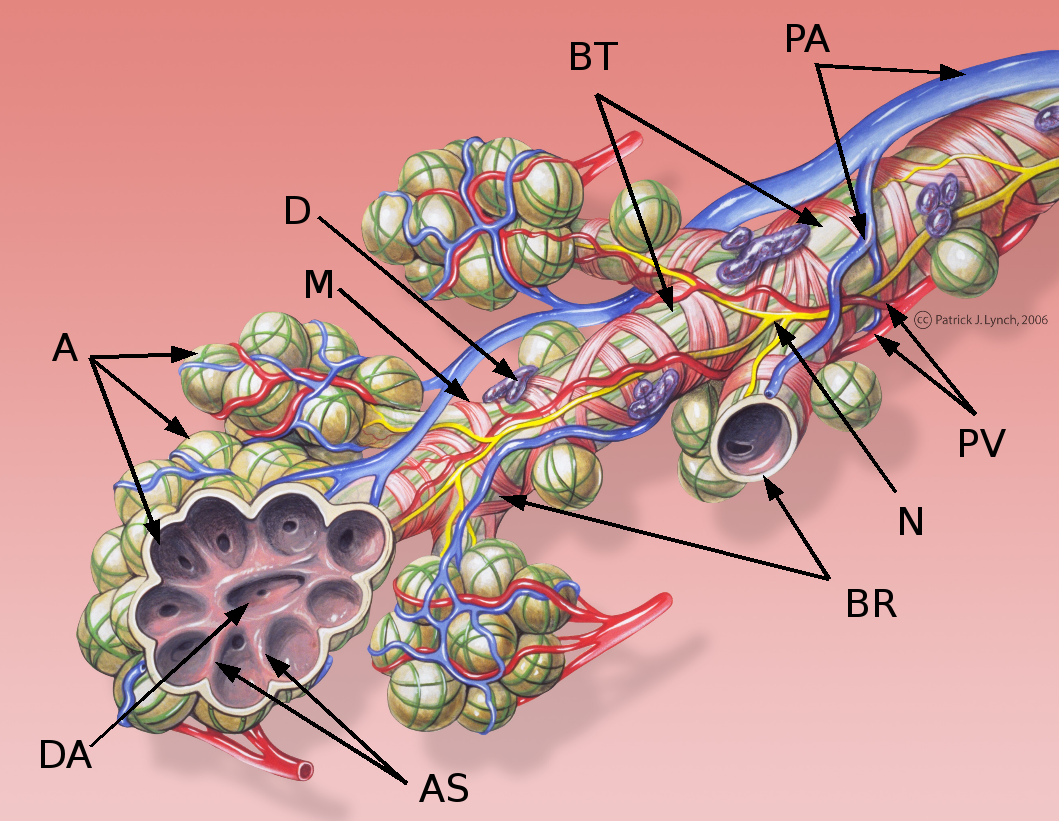
| Description | Bronchial anatomy detail of alveoli and lung circulation |
| Date | |
| Source | Patrick J. Lynch, medical illustrator |
| Author | Patrick J. Lynch, medical illustrator, modified by Christian2003 |
| Permission (Reusing this file) |
Creative Commons Attribution 2.5 License 2006 |
| Other versions | Image:Bronchial anatomy.jpg |
- AS - Septum alveolare
- BR - Bronchus respiratorius
- BT - Bronchus terminalis
- D - Mucous gland
- DA - Ductus alveolaris
- M - Musculus
- N - Nervus
- PA - Branch of Arteria pulm.
- PV - Branch of Vena pulm.
Patrick J. Lynch; illustrator; C. Carl Jaffe; MD; cardiologist Yale University Center for Advanced Instructional Media Medical Illustrations by Patrick Lynch, generated for multimedia teaching projects by the Yale University School of Medicine, Center for Advanced Instructional Media, 1987-2000. Patrick J. Lynch, http://patricklynch.net Creative Commons Attribution 2.5 License 2006; no usage restrictions except please preserve our creative credits: Patrick J. Lynch, medical illustrator; C. Carl Jaffe, MD, cardiologist. https://creativecommons.org/licenses/by/2.5/
Licensing
- You are free:
- to share – to copy, distribute and transmit the work
- to remix – to adapt the work
- Under the following conditions:
- attribution – You must give appropriate credit, provide a link to the license, and indicate if changes were made. You may do so in any reasonable manner, but not in any way that suggests the licensor endorses you or your use.
File history
Click on a date/time to view the file as it appeared at that time.
| Date/Time | Thumbnail | Dimensions | User | Comment | |
|---|---|---|---|---|---|
| current | 11:53, 9 September 2007 | | 1,059 × 821 (817 KB) | Christian2003~commonswiki | == Summary == {{Information |Description = Bronchial anatomy detail of alveoli and lung circulation |Source = Patrick J. Lynch, medical illustrator |Date = December 23, 2006 |Author = Patrick J. Lynch, medical illustrator, modified by [[User:Christian2003 |
File usage
Global file usage
The following other wikis use this file:
- Usage on ar.wikipedia.org
- Usage on azb.wikipedia.org
- Usage on az.wikipedia.org
- Usage on bg.wikipedia.org
- Usage on de.wikipedia.org
- Usage on es.wikipedia.org
- Usage on fa.wikipedia.org
- Usage on fr.wikipedia.org
- Usage on gl.wikipedia.org
- Usage on hy.wikipedia.org
- Usage on ku.wikipedia.org
- Usage on tr.wikipedia.org
- Usage on uk.wikipedia.org