
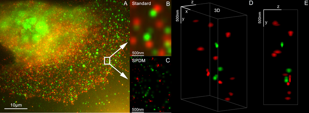
![3D Dual Color Super Resolution Microscopy by combining localization microscopy SPDM with spatially modulated illumination SMI / Optical nanoscopy / Christoph Cremer emeritus at Heidelberg university [1], [2] 3D reconstruction of dual color acquistion of Her2/neu and Her 3. A) conventional wide-field fluorescence image of an AG11132 mammary epithelial cells. Her3 is labelled with Alexa488 (green) and Her2/neu with Alexa568 (red). B) magnified image of a small area of A. C) localization microscopy SPDM of the same region of interest as in B. D) and E) show a 3D reconstruction of the protein clusters using the combination of SPDM and SMI microscopy (LIMON technology). Scale bars in D) and E) are 500 nm in each direction. By combining SPDMphymod with SMI (both invented in Christoph Cremer´s lab in 1996) a 3D dual colour reconstruction of the spatial arrangements of Her2/neu and Her3 clusters was achieved. The positions in all three directions of the protein clusters could be determined with an accuracy of about 25 nm. Her2/neu and Her3 are tyrosine kinase receptors and their overexpression is related to certain types breast cancer. The paper analyses the Her2 distribution pattern in breast cancer cells and counts the clusters (20 637 clusters with a mean diameter of 67 nm) with a specifically developed clusters finding algorithm for super resolution microscopy. This methods could help for the exploration of the resistance mechanism in Trastuzumab/Herceptin treatment. Publication: Rainer Kaufmann, Patrick Müller, Georg Hildenbrand, Michael Hausmann & Christoph Cremer [3], [4] : Analysis of Her2/neu membrane protein clusters in different types of breast cancer cells using localization microscopy, Journal of Microscopy 2010, doi: 10.1111/j.1365-2818.2010.03436.x](http://upload.wikimedia.org/wikipedia/commons/1/1a/3D_Dual_Color_Super_Resolution_Microscopy_Cremer_2010.png)
Original file (3,486 × 1,280 pixels, file size: 3 MB, MIME type: image/png)
| This is a file from the Wikimedia Commons. Information from its description page there is shown below. Commons is a freely licensed media file repository. You can help. |
Summary
| Description |
3D Dual Color Super Resolution Microscopy by combining localization microscopy SPDM with spatially modulated illumination SMI / Optical nanoscopy / Christoph Cremer emeritus at Heidelberg university [1], [2]
3D reconstruction of dual color acquistion of Her2/neu and Her 3. A) conventional wide-field fluorescence image of an AG11132 mammary epithelial cells. Her3 is labelled with Alexa488 (green) and Her2/neu with Alexa568 (red). B) magnified image of a small area of A. C) localization microscopy SPDM of the same region of interest as in B. D) and E) show a 3D reconstruction of the protein clusters using the combination of SPDM and SMI microscopy (LIMON technology). Scale bars in D) and E) are 500 nm in each direction. By combining SPDMphymod with SMI (both invented in Christoph Cremer´s lab in 1996) a 3D dual colour reconstruction of the spatial arrangements of Her2/neu and Her3 clusters was achieved. The positions in all three directions of the protein clusters could be determined with an accuracy of about 25 nm. Her2/neu and Her3 are tyrosine kinase receptors and their overexpression is related to certain types breast cancer. The paper analyses the Her2 distribution pattern in breast cancer cells and counts the clusters (20 637 clusters with a mean diameter of 67 nm) with a specifically developed clusters finding algorithm for super resolution microscopy. This methods could help for the exploration of the resistance mechanism in Trastuzumab/Herceptin treatment. Publication: Rainer Kaufmann, Patrick Müller, Georg Hildenbrand, Michael Hausmann & Christoph Cremer [3], [4] : Analysis of Her2/neu membrane protein clusters in different types of breast cancer cells using localization microscopy, Journal of Microscopy 2010, doi: 10.1111/j.1365-2818.2010.03436.x |
| Date | 051611 |
| Source | Own work |
| Author | Andy Nestl |
Gallery
- Super Resolution Microscopy - Localisation Microscopy
-
 Breast Cancer Cells: 3D Dual Color Super Resolution Microscopy of Her2 and Her3 & cluster calculations
 -
 Single YFP molecule detection in a human cancer cell. Typical distance measurements 15 nm
 -
 Co- localisation microscopy with GFP and RFP fusion proteins (nucleus of a bone cancer cell) 120.000 localized molecules in a widefield area(470 µm2)
 -
 Label-free Localisation Microscopy SPDM - Super Resolution Microscopy reveals prior undetebable intracellular structures
 -
 Investigation of human eye tissue, affected by macular degeneration AMD
 -
 Virus Super Resolution Microscopy SPDM Cremer/Wege labs


Licensing

|
Permission is granted to copy, distribute and/or modify this document under the terms of the GNU Free Documentation License, Version 1.2 or any later version published by the Free Software Foundation; with no Invariant Sections, no Front-Cover Texts, and no Back-Cover Texts. A copy of the license is included in the section entitled GNU Free Documentation License. |
- You are free:
- to share – to copy, distribute and transmit the work
- to remix – to adapt the work
- Under the following conditions:
- attribution – You must give appropriate credit, provide a link to the license, and indicate if changes were made. You may do so in any reasonable manner, but not in any way that suggests the licensor endorses you or your use.
- share alike – If you remix, transform, or build upon the material, you must distribute your contributions under the same or compatible license as the original.
- ↑ https://www.physik.uni-heidelberg.de/personen/lsf.php?details=1537 |titel=Fakultät für Physik und Astronomie |abruf=2020-10-01
- ↑ https://www.kip.uni-heidelberg.de/people/emeriti.php?action=details&num=14
- ↑ http://www.kip.uni-heidelberg.de/AG_Cremer/de
- ↑ https://www.imb.de/research-at-imb/cremer/research/
File history
Click on a date/time to view the file as it appeared at that time.
| Date/Time | Thumbnail | Dimensions | User | Comment | |
|---|---|---|---|---|---|
| current | 12:32, 16 May 2011 | 3,486 × 1,280 (3 MB) | Andy Nestl | {{Information |Description ={{en|1=3D Dual Color Super Resolution Microscopy by combining localization microscopy SPDM with spatially modulated illumination SMI 3D reconstruction of dual color acquistion of Her2/neu and Her 3. A) conventional wide-fie |
File usage
Global file usage
The following other wikis use this file:
- Usage on ar.wikipedia.org
- Usage on bs.wikipedia.org
- Usage on ceb.wikipedia.org
- Usage on cs.wikipedia.org
- Usage on de.wikipedia.org
- Usage on eo.wikipedia.org
- Usage on fa.wikipedia.org
- Usage on gl.wikipedia.org
- Usage on it.wikipedia.org
- Usage on pt.wikipedia.org
- Usage on vi.wikipedia.org
- Usage on zh.wikipedia.org