To install click the Add extension button. That's it.
The source code for the WIKI 2 extension is being checked by specialists of the Mozilla Foundation, Google, and Apple. You could also do it yourself at any point in time.
How to transfigure the Wikipedia
Would you like Wikipedia to always look as professional and up-to-date? We have created a browser extension. It will enhance any encyclopedic page you visit with the magic of the WIKI 2 technology.
Try it — you can delete it anytime.
Install in 5 seconds
Yep, but later
4,5
Kelly Slayton
Congratulations on this excellent venture… what a great idea!
Alexander Grigorievskiy
I use WIKI 2 every day and almost forgot how the original Wikipedia looks like.
Pluripotent: Embryonic stem cells are able to develop into any type of cell, excepting those of the placenta. Only embryonic stem cells of the morula are totipotent: able to develop into any type of cell, including those of the placenta.
Researchers are currently focusing heavily on the therapeutic potential of embryonic stem cells, with clinical use being the goal for many laboratories.[2] Potential uses include the treatment of diabetes and heart disease.[2] The cells are being studied to be used as clinical therapies, models of genetic disorders, and cellular/DNA repair. However, adverse effects in the research and clinical processes such as tumors and unwanted immune responses have also been reported.[5]
YouTube Encyclopedic
1/5
Views:
494 270
2 361
31 912
11 894
82 374
Embryonic stem cells | Cells | MCAT | Khan Academy
Embryonic Stem Cells
GCSE Biology - What are Stem Cells? Difference Between Embryonic and Adult Stem Cells #11
Embryonic Stem Cells and Somatic Cell Nuclear Transfer
Embryonic Stem Cells & their Controversy (unbiased view)
Transcription
Where we left off after the
meiosis videos is that we had
two gametes.
We had a sperm and an egg.
Let me draw the sperm.
So you had the sperm and
then you had an egg.
Maybe I'll do the egg in
a different color.
That's the egg, and we all
know how this story goes.
The sperm fertilizes the egg.
And a whole cascade of events
start occurring.
The walls of the egg then become
impervious to other
sperm so that only one sperm can
get in, but that's not the
focus of this video.
The focus of this video is how
this fertilized egg develops
once it has become a zygote.
So after it's fertilized, you
remember from the meiosis
videos that each of these were
haploid, or that they had--
oh, I added an extra i there--
that they had half the
contingency of the DNA.
As soon as the sperm fertilizes
this egg, now, all
of a sudden, you have
a diploid zygote.
Let me do that.
So now let me pick
a nice color.
So now you're going to have a
diploid zygote that's going to
have a 2N complement of the DNA
material or kind of the
full complement of what a normal
cell in our human body
would have. So this is diploid,
and it's a zygote,
which is just a fancy way of
saying the fertilized egg.
And it's now ready
to essentially
turn into an organism.
So immediately after
fertilization, this zygote
starts experiencing cleavage.
It's experiencing mitosis,
that's the mechanism, but it
doesn't increase
a lot in size.
So this one right here will then
turn into-- it'll just
split up via mitosis
into two like that.
And, of course, these are each
2N, and then those are going
to split into four like that.
And each of these have the same
exact genetic complement
as that first zygote, and
it keeps splitting.
And this mass of cells, we can
start calling it, this right
here, this is referred
to as the morula.
And actually, it comes from the
word for mulberry because
it looks like a mulberry.
So actually, let me just kind
of simplify things a little
bit because we don't
have to start here.
So we start with a zygote.
This is a fertilized egg.
It just starts duplicating via
mitosis, and you end up with a
ball of cells.
It's often going to be a power
of two, because these cells,
at least in the initial stages
are all duplicating all at
once, and then you
have this morula.
Now, once the morula gets to
about 16 cells or so-- and
we're talking about
four or five days.
This isn't an exact process--
they started differentiating a
little bit, where the outer
cells-- and this kind of turns
into a sphere.
Let me make it a little
bit more sphere like.
So it starts differentiating
between-- let me make some
outer cells.
This would be a cross-section
of it.
It's really going to look
more like a sphere.
That's the outer cells and then
you have your inner cells
on the inside.
These outer cells are called
the trophoblasts.
Let me do it in a
different color.
Let me scroll over.
I don't want to go there.
And then the inner cells, and
this is kind of the crux of
what this video is all
about-- let me scroll
down a little bit.
The inner cells-- pick
a suitable color.
The inner cells right there are
called the embryoblast.
And then what's going to happen
is some fluid's going
to start filling in some
of this gap between the
embryoblast and the trophoblast,
so you're going
to start having some fluid that
comes in there, and so
the morula will eventually
look like this, where the
trophoblast, or the outer
membrane, is kind of this huge
sphere of cells.
And this is all happening as
they keep replicating.
Mitosis is the mechanism, so now
my trophoblast is going to
look like that, and then
my embryoblast is going
to look like this.
Sometimes the embryoblast-- so
this is the embryoblast.
Sometimes it's also called the
inner cell mass, so let me
write that.
And this is what's going to
turn into the organism.
And so, just so you know a
couple of the labels that are
involved here, if we're dealing
with a mammalian
organism, and we are mammals,
we call this thing that the
morula turned into is a zygote,
then a morula, then
the cells of the morula started
to differentiate into
the trophoblast, or kind of the
outside cells, and then
the embryoblast. And then you
have this space that forms
here, and this is just fluid,
and it's called the
blastocoel.
A very non-intuitive spelling
of the coel part of
blastocoel.
But once this is formed, this is
called a blastocyst. That's
the entire thing right here.
Let me scroll down
a little bit.
This whole thing is called the
blastocyst, and this is the
case in humans.
Now, it can be a very confusing
topic, because a lot
of times in a lot of books on
biology, you'll say, hey, you
go from the morula to
the blastula or the
blastosphere stage.
Let me write those words down.
So sometimes you'll say morula,
and you go to blastula.
Sometimes it's called
the blastosphere.
And I want to make it very
clear that these are
essentially the same stages
in development.
These are just for-- you know,
in a lot of books, they'll
start talking about frogs or
tadpoles or things like that,
and this applies to them.
While we're talking about
mammals, especially the ones
that are closely related
to us, the stage is the
blastocyst stage, and the real
differentiator is when people
talk about just blastula
and blastospheres.
There isn't necessarily this
differentiation between these
outermost cells and these
embryonic, or this
embryoblast, or this inner
cell mass here.
But since the focus of this
video is humans, and really
that's where I wanted to start
from, because that's what we
are and that's what's
interesting, we're going to
focus on the blastocyst.
Now, everything I've talked
about in this video, it was
really to get to this point,
because what we have here,
these little green cells that
I drew right here in the
blastocysts, this inner cell
mass, this is what will turn
into the organism.
And you say, OK, Sal, if that's
the organism, what's
all of these purple
cells out here?
This trophoblast out there?
That is going to turn into the
placenta, and I'll do a future
video where in a human, it'll
turn into a placenta.
So let me write that down.
It'll turn into the placenta.
And I'll do a whole future video
about I guess how babies
are born, and I actually learned
a ton about that this
past year because a baby
was born in our house.
But the placenta is really
kind of what the embryo
develops inside of, and it's the
interface, especially in
humans and in mammals, between
the developing fetus and its
mother, so it kind of is the
exchange mechanism that
separates their two systems,
but allows the necessary
functions to go on
between them.
But that's not the focus
of this video.
The focus of this video is the
fact that these cells, which
at this point are-- they've
differentiated themselves away
from the placenta cells, but
they still haven't decided
what they're going to become.
Maybe this cell and its
descendants eventually start
becoming part of the nervous
system, while these cells
right here might become muscle
tissue, while these cells
right here might become
the liver.
These cells right here are
called embryonic stem cells,
and probably the first time in
this video you're hearing a
term that you might recognize.
So if I were to just take one of
these cells, and actually,
just to introduce you to another
term, you know, we
have this zygote.
As soon as it starts dividing,
each of these cells are called
a blastomere.
And you're probably wondering,
Sal, why does this word blast
keep appearing in this kind
of embryology video, these
development videos?
And that comes from the Greek
for spore: blastos.
So the organism is beginning
to spore out or grow.
I won't go into the word origins
of it, but that's
where it comes from and that's
why everything has
this blast in it.
So these are blastomeres.
So when I talk what embryonic
stem cells, I'm talking about
the individual blastomeres
inside of this embryoblast or
inside of this inner
cell mass.
These words are actually
unusually fun to say.
So each of these is an
embryonic stem cell.
Let me write this down
in a vibrant color.
So each of these right here are
embryonic stem cells, and
I wanted to get to this.
And the reason why these are
interesting, and I think you
already know, is that there's
a huge debate around these.
One, these have the potential
to turn into anything, that
they have this plasticity.
That's another word that
you might hear.
Let me write that down,
too: plasticity.
And the word essentially comes
from, you know, like a plastic
can turn into anything else.
When we say that something has
plasticity, we're talking
about its potential
to turn into a lot
of different things.
So the theory is, and there's
already some trials that seem
to substantiate this, especially
in some lower
organisms, that, look, if you
have some damage at some point
in your body-- let me
draw a nerve cell.
Let me say I have a-- I won't
go into the actual mechanics
of a nerve cell, but let's say
that we have some damage at
some point on a nerve cell right
there, and because of
that, someone is paralyzed
or there's some nerve
dysfunction.
We're dealing with multiple
sclerosis or who knows what.
The idea is, look, we have these
cell here that could
turn into anything, and we're
just really understanding how
it knows what to turn into.
It really has to look at its
environment and say, hey, what
are the guys around me doing,
and maybe that's what helps
dictate what it does.
But the idea is you take these
things that could turn to
anything and you put them where
the damage is, you layer
them where the damage is, and
then they can turn into the
cell that they need
to turn into.
So in this case, they would
turn into nerve cells.
They would turn to nerve cells
and repair the damage and
maybe cure the paralysis
for that individual.
So it's a huge, exciting area
of research, and you could
even, in theory, grow
new organs.
If someone needs a kidney
transplant or a heart
transplant, maybe in the future,
we could take a colony
of these embryonic stem cells.
Maybe we can put them in some
type of other creature, or who
knows what, and we can turn it
into a replacement heart or a
replacement kidney.
So there's a huge amount
of excitement about
what these can do.
I mean, they could cure a lot of
formerly uncurable diseases
or provide hope for a
lot of patients who
might otherwise die.
But obviously, there's
a debate here.
And the debate all revolves
around the issue of if you
were to go in here and try to
extract one of these cells,
you're going to kill
this embryo.
You're going to kill this
developing embryo, and that
developing embryo had
the potential to
become a human being.
It's a potential that obviously
has to be in the
right environment, and it has
to have a willing mother and
all of the rest, but it does
have the potential.
And so for those, especially, I
think, in the pro-life camp,
who say, hey, anything that has
a potential to be a human
being, that is life and it
should not be killed.
So people on that side of the
camp, they're against the
destroying of this embryo.
I'm not making this video to
take either side to that
argument, but it's a potential
to turn to a human being.
It's a potential, right?
So obviously, there's a huge
amount of debate, but now, now
you know in this video what
people are talking about when
they say embryonic stem cells.
And obviously, the next question
is, hey, well, why
don't they just call them stem
cells as opposed to embryonic
stem cells?
And that's because in all of our
bodies, you do have what
are called somatic stem cells.
Let me write that down.
Somatic or adults stem cells.
And we all have them.
They're in our bone marrow to
help produce red blood cells,
other parts of our body, but the
problem with somatic stem
cells is they're not as plastic,
which means that they
can't form any type of cell
in the human body.
There's an area of research
where people are actually
maybe trying to make them more
plastic, and if they are able
to take these somatic stem
cells and make them more
plastic, it might maybe kill
the need to have these
embryonic stem cells, although
maybe if they do this too
good, maybe these will have
the potential to turn into
human beings as well,
so that could
become a debatable issue.
But right now, this isn't an
area of debate because, left
to their own devices, a somatic
stem cell or an adult
stem cell won't turn into
a human being, while an
embryonic one, if it is
implanted in a willing mother,
then, of course, it will turn
into a human being.
And I want to make one side
note here, because I don't
want to take any sides on the
debate of-- well, I mean,
facts are facts.
This does have the potential
to turn into a human being,
but it also has the potential
to save millions of lives.
Both of those statements are
facts, and then you can decide
on your own which side of that
argument you'd like to or what
side of that balance you
would like to kind of
put your own opinion.
But there's one thing I want
to talk about that in the
public debate is never
brought up.
So you have this notion of when
you-- to get an embryonic
stem cell line, and when I say
a stem cell line, I mean you
take a couple of stem cells, or
let's say you take one stem
cell, and then you put it in a
Petri dish, and then you allow
it to just duplicate.
So this one turns into two,
those two turn to four.
Then someone could take one of
these and then put it in their
own Petri dish.
These are a stem cell line.
They all came from one unique
embryonic stem cell or what
initially was a blastomere.
So that's what they call
a stem cell line.
So the debate obviously is when
you start an embryonic
stem cell line, you are
destroying an embryo.
But I want to make the point
here that embryos are being
destroyed in other processes,
and namely, in-vitro
fertilization.
And maybe this'll be my next
video: fertilization.
And this is just the notion that
they take a set of eggs
out of a mother.
It's usually a couple that's
having trouble having a child,
and they take a bunch of
eggs out of the mother.
So let's say they take
maybe 10 to 30
eggs out of the mother.
They actually perform a surgery,
take them out of the
ovaries of the mother, and then
they fertilize them with
semen, either it might come
from the father or a sperm
donor, so then all of these
becomes zygotes once they're
fertilized with semen.
So these all become zygotes,
and then they allow them to
develop, and they usually allow
them to develop to the
blastocyst stage.
So eventually all of these
turn into blastocysts.
They have a blastocoel in
the center, which is
this area of fluid.
They have, of course, the
embryo, the inner cell mass in
them, and what they do is they
look at the ones that they
deem are healthier or maybe
the ones that are at least
just not unhealthy, and they'll
take a couple of these
and they'll implant these into
the mother, so all of this is
occurring in a Petri dish.
So maybe these four look good,
so they're going to take these
four, and they're going to
implant these into a mother,
and if all goes well, maybe one
of these will turn into--
will give the couple a child.
So this one will develop and
maybe the other ones won't.
But if you've seen John & Kate
Plus 8, you know that many
times they implant a lot of
them in there, just to
increase the probability that
you get at least one child.
But every now and then, they
implant seven or eight, and
then you end up with
eight kids.
And that's why in-vitro
fertilization often results in
kind of these multiple
births, or reality
television shows on cable.
But what do they do with all
of these other perfectly--
well, I won't say perfectly
viable, but these are embryos.
They may or may not be perfectly
viable, but you have
these embryos that have the
potential, just like this one
right here.
These all have the potential
to turn into a human being.
But most fertility clinics,
roughly half of them, they
either throw these away,
they destroy them, they
allow them to die.
A lot of these are frozen, but
just the process of freezing
them kills them and then bonding
them kills them again,
so most of these, the process of
in-vitro fertilization, for
every one child that has the
potential to develop into a
full-fledged human being, you're
actually destroying
tens of very viable embryos.
So at least my take on it is
if you're against-- and I
generally don't want to take a
side on this, but if you are
against research that involves
embryonic stem cells because
of the destruction of embryos,
on that same, I guess,
philosophical ground, you
should also be against
in-vitro fertilization because
both of these involve the
destruction of zygotes.
I think-- well, I won't talk
more about this, because I
really don't want to take sides,
but I want to show that
there is kind of an equivalence
here that's
completely lost in this debate
on whether embryonic stem
cells should be used because
they have a destruction of
embryos, because you're
destroying just as many
embryos in this-- well, I won't
say just as many, but
you are destroying embryos.
There's hundreds of thousands of
embryos that get destroyed
and get frozen and obviously
destroyed in that process as
well through this in-vitro
fertilization process.
So anyway, now hopefully you
have the tools to kind of
engage in the debate around stem
cells, and you see that
it all comes from what we
learned about meiosis.
They produce these gametes.
The male gamete fertilizes
a female gamete.
The zygote happens or gets
created and starts splitting
up the morula, and then it
keeps splitting and it
differentiates into the
blastocyst, and then this is
where the stem cells are.
So you already know enough
science to engage in kind of a
very heated debate.
Properties
IPS Cell
The transcriptome of embryonic stem cells
Embryonic stem cells (ESCs), derived from the blastocyst stage of early mammalian embryos, are distinguished by their ability to differentiate into any embryonic cell type and by their ability to self-renew. It is these traits that makes them valuable in the scientific and medical fields. ESCs have a normal karyotype, maintain high telomerase activity, and exhibit remarkable long-term proliferative potential.[6]
Pluripotent
Embryonic stem cells of the inner cell mass are pluripotent, meaning they are able to differentiate to generate primitive ectoderm, which ultimately differentiates during gastrulation into all derivatives of the three primary germ layers: ectoderm, endoderm, and mesoderm. These germ layers generate each of the more than 220 cell types in the adult human body. When provided with the appropriate signals, ESCs initially form precursor cells that in subsequently differentiate into the desired cell types. Pluripotency distinguishes embryonic stem cells from adult stem cells, which are multipotent and can only produce a limited number of cell types.
Self renewal and repair of structure
Under defined conditions, embryonic stem cells are capable of self-renewing indefinitely in an undifferentiated state. Self-renewal conditions must prevent the cells from clumping and maintain an environment that supports an unspecialized state.[7] Typically this is done in the lab with media containing serum and leukemia inhibitory factor or serum-free media supplements with two inhibitory drugs ("2i"), the MEK inhibitor PD03259010 and GSK-3 inhibitor CHIR99021.[8]
Growth
ESCs divide very frequently due to a shortened G1 phase in their cell cycle. Rapid cell division allows the cells to quickly grow in number, but not size, which is important for early embryo development. In ESCs, cyclin A and cyclin E proteins involved in the G1/S transition are always expressed at high levels.[9]Cyclin-dependent kinases such as CDK2 that promote cell cycle progression are overactive, in part due to downregulation of their inhibitors.[10]Retinoblastoma proteins that inhibit the transcription factorE2F until the cell is ready to enter S phase are hyperphosphorylated and inactivated in ESCs, leading to continual expression of proliferation genes.[9] These changes result in accelerated cycles of cell division. Although high expression levels of pro-proliferative proteins and a shortened G1 phase have been linked to maintenance of pluripotency,[11][12] ESCs grown in serum-free 2i conditions do express hypo-phosphorylated active Retinoblastoma proteins and have an elongated G1 phase.[13] Despite this difference in the cell cycle when compared to ESCs grown in media containing serum these cells have similar pluripotent characteristics.[14] Pluripotency factors Oct4 and Nanog play a role in transcriptionally regulating the embryonic stem cell cycle.[15][16]
Uses
Due to their plasticity and potentially unlimited capacity for self-renewal, embryonic stem cell therapies have been proposed for regenerative medicine and tissue replacement after injury or disease. Pluripotent stem cells have shown promise in treating a number of varying conditions, including but not limited to: spinal cord injuries, age related macular degeneration, diabetes, neurodegenerative disorders (such as Parkinson's disease), AIDS, etc.[17] In addition to their potential in regenerative medicine, embryonic stem cells provide a possible alternative source of tissue/organs which serves as a possible solution to the donor shortage dilemma. There are some ethical controversies surrounding this though (see Ethical debate section below). Aside from these uses, ESCs can also be used for research on early human development, certain genetic disease, and in vitrotoxicology testing.[6]
Utilizations
According to a 2002 article in PNAS, "Human embryonic stem cells have the potential to differentiate into various cell types, and, thus, may be useful as a source of cells for transplantation or tissue engineering."[18]
Tissue engineering
Embryoid bodies 24 hours after formation.
In tissue engineering, the use of stem cells are known to be of importance. In order to successfully engineer a tissue, the cells used must be able to perform specific biological functions such as secretion of cytokines, signaling molecules, interacting with neighboring cells, and producing an extracellular matrix in the correct organization. Stem cells demonstrates these specific biological functions along with being able to self-renew and differentiate into one or more types of specialized cells. Embryonic stem cells is one of the sources that are being considered for the use of tissue engineering.[19] The use of human embryonic stem cells have opened many new possibilities for tissue engineering, however, there are many hurdles that must be made before human embryonic stem cell can even be utilized. It is theorized that if embryonic stem cells can be altered to not evoke the immune response when implanted into the patient then this would be a revolutionary step in tissue engineering.[20] Embryonic stem cells are not limited to tissue engineering.
Cell replacement therapies
Research has focused on differentiating ESCs into a variety of cell types for eventual use as cell replacement therapies. Some of the cell types that have or are currently being developed include cardiomyocytes, neurons, hepatocytes, bone marrow cells, islet cells and endothelial cells.[21] However, the derivation of such cell types from ESCs is not without obstacles, therefore research has focused on overcoming these barriers. For example, studies are underway to differentiate ESCs into tissue specific cardiomyocytes and to eradicate their immature properties that distinguish them from adult cardiomyocytes.[22]
Clinical potential
Researchers have differentiated ESCs into dopamine-producing cells with the hope that these neurons could be used in the treatment of Parkinson's disease.[23][24]
Studies involving ESCs are underway to provide an alternative treatment for diabetes. For example ESCs have been differentiated into insulin-producing cells,[26] and researchers at Harvard University were able to produce large quantities of pancreatic beta cells from ESCs.[27]
An article published in the European Heart Journal describes a translational process of generating human embryonic stem cell-derived cardiac progenitor cells to be used in clinical trials of patients with severe heart failure.[28]
Drug discovery
Besides becoming an important alternative to organ transplants, ESCs are also being used in the field of toxicology, and as cellular screens to uncover new chemical entities that can be developed as small-molecule drugs. Studies have shown that cardiomyocytes derived from ESCs are validated in vitro models to test drug responses and predict toxicity profiles.[21] ESC derived cardiomyocytes have been shown to respond to pharmacological stimuli and hence can be used to assess cardiotoxicity such as torsades de pointes.[29]
ESC-derived hepatocytes are also useful models that could be used in the preclinical stages of drug discovery. However, the development of hepatocytes from ESCs has proven to be challenging and this hinders the ability to test drug metabolism. Therefore, research has focused on establishing fully functional ESC-derived hepatocytes with stable phase I and II enzyme activity.[30]
Models of genetic disorder
Several new studies have started to address the concept of modeling genetic disorders with embryonic stem cells. Either by genetically manipulating the cells, or more recently, by deriving diseased cell lines identified by prenatal genetic diagnosis (PGD), modeling genetic disorders is something that has been accomplished with stem cells. This approach may very well prove valuable at studying disorders such as Fragile-X syndrome, Cystic fibrosis, and other genetic maladies that have no reliable model system.
Yury Verlinsky, a Russian-American medical researcher who specialized in embryo and cellular genetics (genetic cytology), developed prenatal diagnosis testing methods to determine genetic and chromosomal disorders a month and a half earlier than standard amniocentesis. The techniques are now used by many pregnant women and prospective parents, especially couples who have a history of genetic abnormalities or where the woman is over the age of 35 (when the risk of genetically related disorders is higher). In addition, by allowing parents to select an embryo without genetic disorders, they have the potential of saving the lives of siblings that already had similar disorders and diseases using cells from the disease free offspring.[31]
Repair of DNA damage
Differentiated somatic cells and ES cells use different strategies for dealing with DNA damage. For instance, human foreskin fibroblasts, one type of somatic cell, use non-homologous end joining (NHEJ), an error prone DNA repair process, as the primary pathway for repairing double-strand breaks (DSBs) during all cell cycle stages.[32] Because of its error-prone nature, NHEJ tends to produce mutations in a cell's clonal descendants.
ES cells use a different strategy to deal with DSBs.[33] Because ES cells give rise to all of the cell types of an organism including the cells of the germ line, mutations arising in ES cells due to faulty DNA repair are a more serious problem than in differentiated somatic cells. Consequently, robust mechanisms are needed in ES cells to repair DNA damages accurately, and if repair fails, to remove those cells with un-repaired DNA damages. Thus, mouse ES cells predominantly use high fidelity homologous recombinational repair (HRR) to repair DSBs.[33] This type of repair depends on the interaction of the two sister chromosomes[verification needed] formed during S phase and present together during the G2 phase of the cell cycle. HRR can accurately repair DSBs in one sister chromosome by using intact information from the other sister chromosome. Cells in the G1 phase of the cell cycle (i.e. after metaphase/cell division but prior the next round of replication) have only one copy of each chromosome (i.e. sister chromosomes aren't present). Mouse ES cells lack a G1 checkpoint and do not undergo cell cycle arrest upon acquiring DNA damage.[34] Rather they undergo programmed cell death (apoptosis) in response to DNA damage.[35] Apoptosis can be used as a fail-safe strategy to remove cells with un-repaired DNA damages in order to avoid mutation and progression to cancer.[36] Consistent with this strategy, mouse ES stem cells have a mutation frequency about 100-fold lower than that of isogenic mouse somatic cells.[37]
On January 23, 2009, Phase I clinical trials for transplantation of oligodendrocytes (a cell type of the brain and spinal cord) derived from human ESCs into spinal cord-injured individuals received approval from the U.S. Food and Drug Administration (FDA), marking it the world's first human ESC human trial.[38] The study leading to this scientific advancement was conducted by Hans Keirstead and colleagues at the University of California, Irvine and supported by Geron Corporation of Menlo Park, CA, founded by Michael D. West, PhD. A previous experiment had shown an improvement in locomotor recovery in spinal cord-injured rats after a 7-day delayed transplantation of human ESCs that had been pushed into an oligodendrocytic lineage.[39] The phase I clinical study was designed to enroll about eight to ten paraplegics who have had their injuries no longer than two weeks before the trial begins, since the cells must be injected before scar tissue is able to form. The researchers emphasized that the injections were not expected to fully cure the patients and restore all mobility. Based on the results of the rodent trials, researchers speculated that restoration of myelin sheathes and an increase in mobility might occur. This first trial was primarily designed to test the safety of these procedures and if everything went well, it was hoped that it would lead to future studies that involve people with more severe disabilities.[40] The trial was put on hold in August 2009 due to FDA concerns regarding a small number of microscopic cysts found in several treated rat models but the hold was lifted on July 30, 2010.[41]
In October 2010 researchers enrolled and administered ESCs to the first patient at Shepherd Center in Atlanta.[42] The makers of the stem cell therapy, Geron Corporation, estimated that it would take several months for the stem cells to replicate and for the GRNOPC1 therapy to be evaluated for success or failure.
In November 2011 Geron announced it was halting the trial and dropping out of stem cell research for financial reasons, but would continue to monitor existing patients, and was attempting to find a partner that could continue their research.[43] In 2013 BioTime, led by CEO Dr. Michael D. West, acquired all of Geron's stem cell assets, with the stated intention of restarting Geron's embryonic stem cell-based clinical trial for spinal cord injury research.[44]
BioTime company Asterias Biotherapeutics (NYSE MKT: AST) was granted a $14.3 million Strategic Partnership Award by the California Institute for Regenerative Medicine (CIRM) to re-initiate the world's first embryonic stem cell-based human clinical trial, for spinal cord injury. Supported by California public funds, CIRM is the largest funder of stem cell-related research and development in the world.[45]
The award provides funding for Asterias to reinitiate clinical development of AST-OPC1 in subjects with spinal cord injury and to expand clinical testing of escalating doses in the target population intended for future pivotal trials.[45]
AST-OPC1 is a population of cells derived from human embryonic stem cells (hESCs) that contains oligodendrocyte progenitor cells (OPCs). OPCs and their mature derivatives called oligodendrocytes provide critical functional support for nerve cells in the spinal cord and brain. Asterias recently presented the results from phase 1 clinical trial testing of a low dose of AST-OPC1 in patients with neurologically complete thoracic spinal cord injury. The results showed that AST-OPC1 was successfully delivered to the injured spinal cord site. Patients followed 2–3 years after AST-OPC1 administration showed no evidence of serious adverse events associated with the cells in detailed follow-up assessments including frequent neurological exams and MRIs. Immune monitoring of subjects through one year post-transplantation showed no evidence of antibody-based or cellular immune responses to AST-OPC1. In four of the five subjects, serial MRI scans performed throughout the 2–3 year follow-up period indicate that reduced spinal cord cavitation may have occurred and that AST-OPC1 may have had some positive effects in reducing spinal cord tissue deterioration. There was no unexpected neurological degeneration or improvement in the five subjects in the trial as evaluated by the International Standards for Neurological Classification of Spinal Cord Injury (ISNCSCI) exam.[45]
The Strategic Partnership III grant from CIRM will provide funding to Asterias to support the next clinical trial of AST-OPC1 in subjects with spinal cord injury, and for Asterias' product development efforts to refine and scale manufacturing methods to support later-stage trials and eventually commercialization. CIRM funding will be conditional on FDA approval for the trial, completion of a definitive agreement between Asterias and CIRM, and Asterias' continued progress toward the achievement of certain pre-defined project milestones.[45]
Concern and controversy
Adverse effects
The major concern with the possible transplantation of ESCs into patients as therapies is their ability to form tumors including teratomas.[46] Safety issues prompted the FDA to place a hold on the first ESC clinical trial, however no tumors were observed.
The main strategy to enhance the safety of ESCs for potential clinical use is to differentiate the ESCs into specific cell types (e.g. neurons, muscle, liver cells) that have reduced or eliminated ability to cause tumors. Following differentiation, the cells are subjected to sorting by flow cytometry for further purification. ESCs are predicted to be inherently safer than iPS cells created with genetically integrating viral vectors because they are not genetically modified with genes such as c-Myc that are linked to cancer. Nonetheless, ESCs express very high levels of the iPS inducing genes and these genes including Myc are essential for ESC self-renewal and pluripotency,[47] and potential strategies to improve safety by eliminating c-Myc expression are unlikely to preserve the cells' "stemness". However, N-myc and L-myc have been identified to induce iPS cells instead of c-myc with similar efficiency.[48] Later protocols to induce pluripotency bypass these problems completely by using non-integrating RNA viral vectors such as sendai virus or mRNA transfection.
Due to the nature of embryonic stem cell research, there are a lot of controversial opinions on the topic. Since harvesting embryonic stem cells usually necessitates destroying the embryo from which those cells are obtained, the moral status of the embryo comes into question. Some people claim that the 5-day-old mass of cells is too young to achieve personhood or that the embryo, if donated from an IVF clinic (where labs typically acquire embryos), would otherwise go to medical waste anyway. Opponents of ESC research claim that an embryo is a human life, therefore destroying it is murder and the embryo must be protected under the same ethical view as a more developed human being.[49]
History
1964: Lewis Kleinsmith and G. Barry Pierce Jr. isolated a single type of cell from a teratocarcinoma, a tumor now known from a germ cell.[50] These cells were isolated from the teratocarcinoma replicated and grew in cell culture as a stem cell and are now known as embryonal carcinoma (EC) cells.[citation needed] Although similarities in morphology and differentiating potential (pluripotency) led to the use of EC cells as the in vitro model for early mouse development,[51] EC cells harbor genetic mutations and often abnormal karyotypes that accumulated during the development of the teratocarcinoma. These genetic aberrations further emphasized the need to be able to culture pluripotent cells directly from the inner cell mass.
Martin Evans revealed a new technique for culturing the mouse embryos in the uterus to allow for the derivation of ES cells from these embryos.
1981: Embryonic stem cells (ES cells) were independently first derived from a mouse embryos by two groups. Martin Evans and Matthew Kaufman from the Department of Genetics, University of Cambridge published first in July, revealing a new technique for culturing the mouse embryos in the uterus to allow for an increase in cell number, allowing for the derivation of ES cell from these embryos.[52]Gail R. Martin, from the Department of Anatomy, University of California, San Francisco, published her paper in December and coined the term "Embryonic Stem Cell".[53] She showed that embryos could be cultured in vitro and that ES cells could be derived from these embryos.
1989: Mario R. Cappechi, Martin J. Evans, and Oliver Smithies publish their research that details their isolation and genetic modifications of embryonic stem cells, creating the first "knockout mice".[54] In creating knockout mice, this publication provided scientists with an entirely new way to study disease.
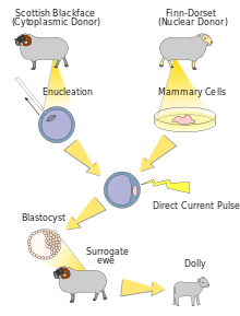
Dolly the sheep cell differentiation1996: Dolly, was the first mammal cloned from an adult cell by the Roslin Institute of the University of Edinburgh.[55] This experiment instituted the proposition that specialized adult cells obtain the genetic makeup to perform a specific task; which established a basis for further research within a variety of cloning techniques. The Dolly experiment was performed by obtaining the mammalian udder cells from a sheep (Dolly) and differentiating these cells until division was concluded. An egg cell was then procured from a different sheep host and the nucleus was removed. An udder cell was placed next to the egg cell and connected by electricity causing this cell to share DNA. This egg cell differentiated into an embryo and the embryo was inserted into a third sheep which gave birth to the clone version of Dolly.[56]
1998: A team from the University of Wisconsin, Madison (James A. Thomson, Joseph Itskovitz-Eldor, Sander S. Shapiro, Michelle A. Waknitz, Jennifer J. Swiergiel, Vivienne S. Marshall, and Jeffrey M. Jones) publish a paper titled "Embryonic Stem Cell Lines Derived From Human Blastocysts". The researchers behind this study not only created the first embryonic stem cells, but recognized their pluripotency, as well as their capacity for self-renewal. The abstract of the paper notes the significance of the discovery with regards to the fields of developmental biology and drug discovery.[57]
2001: President George W. Bush allows federal funding to support research on roughly 60—at this time, already existing—lines of embryonic stem cells. Seeing as the limited lines that Bush allowed research on had already been established, this law supported embryonic stem cell research without raising any ethical questions that could arise with the creation of new lines under federal budget.[58]
2006: Japanese scientists Shinya Yamanaka and Kazutoshi Takashi publish a paper describing the induction of pluripotent stem cells from cultures of adult mouse fibroblasts. Induced pluripotent stem cells (iPSCs) are a huge discovery, as they are seemingly identical to embryonic stem cells and could be used without sparking the same moral controversy.[59]
January, 2009: The US Food and Drug Administration (FDA) provides approval for Geron Corporation's phase I trial of their human embryonic stem cell-derived treatment for spinal cord injuries. The announcement was met with excitement from the scientific community, but also with wariness from stem cell opposers. The treatment cells were, however, derived from the cell lines approved under George W. Bush's ESC policy.[60]
March, 2009: Executive Order 13505 is signed by President Barack Obama, removing the restrictions put in place on federal funding for human stem cells by the previous presidential administration. This would allow the National Institutes of Health (NIH) to provide funding for hESC research. The document also states that the NIH must provide revised federal funding guidelines within 120 days of the order's signing.[61]
Techniques and conditions for derivation and culture
Derivation from humans
In vitro fertilization generates multiple embryos. The surplus of embryos is not clinically used or is unsuitable for implantation into the patient, and therefore may be donated by the donor with consent. Human embryonic stem cells can be derived from these donated embryos or additionally they can also be extracted from cloned embryos created using a cell from a patient and a donated egg through the process of somatic cell nuclear transfer.[62] The inner cell mass (cells of interest), from the blastocyst stage of the embryo, is separated from the trophectoderm, the cells that would differentiate into extra-embryonic tissue. Immunosurgery, the process in which antibodies are bound to the trophectoderm and removed by another solution, and mechanical dissection are performed to achieve separation. The resulting inner cell mass cells are plated onto cells that will supply support. The inner cell mass cells attach and expand further to form a human embryonic cell line, which are undifferentiated. These cells are fed daily and are enzymatically or mechanically separated every four to seven days. For differentiation to occur, the human embryonic stem cell line is removed from the supporting cells to form embryoid bodies, is co-cultured with a serum containing necessary signals, or is grafted in a three-dimensional scaffold to result.[63]
Derivation from other animals
Embryonic stem cells are derived from the inner cell mass of the early embryo, which are harvested from the donor mother animal. Martin Evans and Matthew Kaufman reported a technique that delays embryo implantation, allowing the inner cell mass to increase. This process includes removing the donor mother's ovaries and dosing her with progesterone, changing the hormone environment, which causes the embryos to remain free in the uterus. After 4–6 days of this intrauterine culture, the embryos are harvested and grown in in vitro culture until the inner cell mass forms “egg cylinder-like structures,” which are dissociated into single cells, and plated on fibroblasts treated with mitomycin-c (to prevent fibroblast mitosis). Clonal cell lines are created by growing up a single cell. Evans and Kaufman showed that the cells grown out from these cultures could form teratomas and embryoid bodies, and differentiate in vitro, all of which indicating that the cells are pluripotent.[52]
Gail Martin derived and cultured her ES cells differently. She removed the embryos from the donor mother at approximately 76 hours after copulation and cultured them overnight in a medium containing serum. The following day, she removed the inner cell mass from the late blastocyst using microsurgery. The extracted inner cell mass was cultured on fibroblasts treated with mitomycin-c in a medium containing serum and conditioned by ES cells. After approximately one week, colonies of cells grew out. These cells grew in culture and demonstrated pluripotent characteristics, as demonstrated by the ability to form teratomas, differentiate in vitro, and form embryoid bodies. Martin referred to these cells as ES cells.[53]
It is now known that the feeder cells provide leukemia inhibitory factor (LIF) and serum provides bone morphogenetic proteins (BMPs) that are necessary to prevent ES cells from differentiating.[64][65] These factors are extremely important for the efficiency of deriving ES cells. Furthermore, it has been demonstrated that different mouse strains have different efficiencies for isolating ES cells.[66] Current uses for mouse ES cells include the generation of transgenic mice, including knockout mice. For human treatment, there is a need for patient specific pluripotent cells. Generation of human ES cells is more difficult and faces ethical issues. So, in addition to human ES cell research, many groups are focused on the generation of induced pluripotent stem cells (iPS cells).[67]
Potential methods for new cell line derivation
On August 23, 2006, the online edition of Nature scientific journal published a letter by Dr. Robert Lanza (medical director of Advanced Cell Technology in Worcester, MA) stating that his team had found a way to extract embryonic stem cells without destroying the actual embryo.[68] This technical achievement would potentially enable scientists to work with new lines of embryonic stem cells derived using public funding in the US, where federal funding was at the time limited to research using embryonic stem cell lines derived prior to August 2001. In March, 2009, the limitation was lifted.[69]
Human embryonic stem cells have also been derived by somatic cell nuclear transfer (SCNT).[70][71] This approach has also sometimes been referred to as "therapeutic cloning" because SCNT bears similarity to other kinds of cloning in that nuclei are transferred from a somatic cell into an enucleated zygote. However, in this case SCNT was used to produce embryonic stem cell lines in a lab, not living organisms via a pregnancy. The "therapeutic" part of the name is included because of the hope that SCNT produced embryonic stem cells could have clinical utility.
The iPS cell technology was pioneered by Shinya Yamanaka's lab in Kyoto, Japan, who showed in 2006 that the introduction of four specific genes encoding transcription factors could convert adult cells into pluripotent stem cells.[72] He was awarded the 2012 Nobel Prize along with Sir John Gurdon "for the discovery that mature cells can be reprogrammed to become pluripotent."[73]
In 2007, it was shown that pluripotentstem cells, highly similar to embryonic stem cells, can be induced by the delivery of four factors (Oct3/4, Sox2, c-Myc, and Klf4) to differentiated cells.[74] Utilizing the four genes previously listed, the differentiated cells are "reprogrammed" into pluripotent stem cells, allowing for the generation of pluripotent/embryonic stem cells without the embryo. The morphology and growth factors of these lab induced pluripotent cells, are equivalent to embryonic stem cells, leading these cells to be known as induced pluripotent stem cells (iPS cells).[75] This observation was observed in mouse pluripotent stem cells, originally, but now can be performed in human adult fibroblasts using the same four genes. [76]
Because ethical concerns regarding embryonic stem cells typically are about their derivation from terminated embryos, it is believed that reprogramming to these iPS cells may be less controversial.
This may enable the generation of patient specific ES cell lines that could potentially be used for cell replacement therapies. In addition, this will allow the generation of ES cell lines from patients with a variety of genetic diseases and will provide invaluable models to study those diseases.
On January 16, 2008, a California-based company, Stemagen, announced that they had created the first mature cloned human embryos from single skin cells taken from adults. These embryos can be harvested for patient matching embryonic stem cells.[79]
Contamination by reagents used in cell culture
The online edition of Nature Medicine published a study on January 24, 2005, which stated that the human embryonic stem cells available for federally funded research are contaminated with non-human molecules from the culture medium used to grow the cells.[80] It is a common technique to use mouse cells and other animal cells to maintain the pluripotency of actively dividing stem cells. The problem was discovered when non-human sialic acid in the growth medium was found to compromise the potential uses of the embryonic stem cells in humans, according to scientists at the University of California, San Diego.[81]
However, a study published in the online edition of Lancet Medical Journal on March 8, 2005, detailed information about a new stem cell line that was derived from human embryos under completely cell- and serum-free conditions. After more than 6 months of undifferentiated proliferation, these cells demonstrated the potential to form derivatives of all three embryonic germ layers both in vitro and in teratomas. These properties were also successfully maintained (for more than 30 passages) with the established stem cell lines.[82]
Muse cells (Multi-lineage differentiating stress enduring cell) are non-cancerouspluripotent stem cell found in adults.[83][84] They were discovered in 2010 by Mari Dezawa and her research group.[83] Muse cells reside in the connective tissue of nearly every organ including the umbilical cord, bone marrow and peripheral blood.[85][83][86][87][88] They are collectable from commercially obtainable mesenchymal cells such as human fibroblasts, bone marrow-mesenchymal stem cells and adipose-derived stem cells.[89][90][91] Muse cells are able to generate cells representative of all three germ layers from a single cell both spontaneously and under cytokine induction. Expression of pluripotency genes and triploblastic differentiation are self-renewable over generations. Muse cells do not undergo teratoma formation when transplanted into a host environment in vivo, eradicating the risk of tumorigenesis through unbridled cell proliferation.[83]
^Bernstein C, Bernstein H, Payne CM, Garewal H (June 2002). "DNA repair/pro-apoptotic dual-role proteins in five major DNA repair pathways: fail-safe protection against carcinogenesis". Mutat. Res. 511 (2): 145–178. doi:10.1016/S1383-5742(02)00009-1. PMID12052432.
^Klotzko, Arlene Judith; Klotzko, Visiting Scholar Royal Free and University College Medical School Arlene Judith (2006). A Clone of Your Own?. Cambridge University Press. ISBN978-0-521-85294-4.
^Ledermann B, Bürki K (1991). "Establishment of a germ-line competent C57BL/6 embryonic stem cell line". Exp Cell Res. 197 (2): 254–258. doi:10.1016/0014-4827(91)90430-3. PMID1959560.
^Takahashi K, Tanabe K, Ohnuki M, Narita M, Ichisaka T, Tomoda K, Yamanaka S (2007). "Induction of pluripotent stem cells from adult human fibroblasts by defined factors". Cell. 131 (5): 861–872. doi:10.1016/j.cell.2007.11.019. hdl:2433/49782. PMID18035408. S2CID8531539.
^Zikuan Leng 1 2, Dongming Sun 2, Zihao Huang 3, Iman Tadmori 2, Ning Chiang 2, Nikhit Kethidi 2, Ahmed Sabra 2, Yoshihiro Kushida 4, Yu-Show Fu 3, Mari Dezawa 4, Xijing He 1, Wise Young 2Quantitative Analysis of SSEA3+ Cells from Human Umbilical Cord after Magnetic SortingCell Transplant
. 2019 Jul;28(7):907–923.