
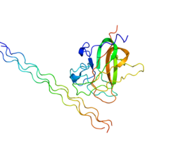


Discoidin domain-containing receptor 2, also known as CD167b (cluster of differentiation 167b), is a protein that in humans is encoded by the DDR2 gene.[5] Discoidin domain-containing receptor 2 is a receptor tyrosine kinase (RTK).
YouTube Encyclopedic
-
1/4Views:447 07610 281506578
-
Cell Junctions | Cells | MCAT | Khan Academy
-
Matrix metalloproteinases
-
Lecture 3: Fluid Mosaic Membrane Model (Cell Biology): Zoology UPSC, M.Sc, B.Sc
-
Mechanisms of Obesity and Leptin in Breast Cancer
Transcription
Voiceover: In this video we're gonna talk a little bit about cell junctions. Cell junctions are basically things that connect cells to other cells. And they often occur in epithelial tissue. We're gonna talk about three major types of cell junctions today. The first, tight junctions, the second desmosomes, and the third, gap junctions. So starting off with tight junctions. Let's say we have two cells like this. So tight junctions basically connect the two cells together. And it's kinda like a glue that connects them together really tightly like that. I think of it like a watertight seal. It's a complete fluid barrier, which means that if there's water or ions or other molecules trying to get through between the two cells, they would not be able to. So it blocks out pretty much everything, from both sides of the cells, so water and ions cannot go through that gap between the two cells. This tight junction, this watertight seal tends to occur in things like the bladder, for example. Or sometimes intestines and the kidney. They occur in places where water really cannot go to other places. For example, our bladder holds urine, which is waste, and it would be really bad for our body if our bladder was unable to be watertight, to hold that urine just within the bladder. The next type that we're gonna talk about are desmosomes. Now let's say we have again, two cells like this. What desmosomes do, is I'm exaggerating the gap between these a little bit, but they're kinda like connections that hold two cells together. And these connections actually attach inside the cytoskeleton. And again, this gap is exaggerated. Usually the cells are a little closer. But in desmosomes, if there is water or ions, they can actually flow between these cells. So ions like sodium or potassium or water, or other small molecules can actually come through in this gap. I like to think about desmosomes kinda like spot welds They kinda hold the two cells together, but it's not like a complete glued seal like the tight junctions. They're kind of spotted throughout the cell so that things can actually flow in between the cells. Desomosomes tend to occur in tissues that experience a lot of stress. They offer a little bit of space for stress relief. So these spot welds, these desmosomes can be found in our skin and in our intestines. Now you notice that our intestines actually have both desmosomes and tight junctions. These cell junctions can be scattered throughout the even the same type of cell. So intestinal tissue can have both tight junctions and desmosomes. The last one we're gonna talk about are gap junctions. So let's say we have, again, our two cells like this. Gap junctions, and again I'm exaggerating the size of our gap junction. But, they're kinda like a tunnel that actually exists between the cells. So they're like a tunnel. And what they do is they actually let water and ions and so on, flow through this gap between the two cells. So they're kinda like a tunnel. These are often found in cells or tissue that spread action potential, or cells that use electrical coupling. For example, they can be found in cardiac muscle. This allows our cardiac muscle to actually spread action potential by using these ions. This allows our heart to continue beating. It can also be found in neurons. So in summary, we have three main types of cell junctions. The first are tight junctions. These are a watertight seal which prevents water or ions from flowing in between cells. We have desmosomes which are spot welds. And these spot welds generally occur in areas of stress, and they also allow water, ions, and other small molecules to flow between cells. And lastly are gap junctions. And gap junctions are tunnels that kind of connect two cells. And these tend to occur in cells that require propagation of electrical signal.
Function
RTKs play a key role in the communication of cells with their microenvironment. These molecules are involved in the regulation of cell growth, differentiation, and metabolism. In several cases the biochemical mechanism by which RTKs transduce signals across the membrane has been shown to be ligand induced receptor oligomerization and subsequent intracellular phosphorylation. In the case of DDR2, the ligand is collagen which binds to its extracellular discoidin domain.[6] This autophosphorylation leads to phosphorylation of cytosolic targets as well as association with other molecules, which are involved in pleiotropic effects of signal transduction. DDR2 has been associated with a number of diseases including fibrosis and cancer.[7]
Structure
RTKs have a tripartite structure with extracellular, transmembrane, and cytoplasmic regions. This gene encodes a member of a novel subclass of RTKs and contains a distinct extracellular region encompassing a factor VIII-like domain.[5]
Gene
Alternative splicing in the 5' UTR of the DDR2 gene results in multiple transcript variants encoding the same protein.[5]
Interactions
DDR2 (gene) has been shown to interact with SHC1[8] and phosphorylate Shp2.[9] DDR2 also interacts with Integrin α1β1 and α2β1 by promoting their adhesion to collagen.[10]
References
- ^ a b c GRCh38: Ensembl release 89: ENSG00000162733 - Ensembl, May 2017
- ^ a b c GRCm38: Ensembl release 89: ENSMUSG00000026674 - Ensembl, May 2017
- ^ "Human PubMed Reference:". National Center for Biotechnology Information, U.S. National Library of Medicine.
- ^ "Mouse PubMed Reference:". National Center for Biotechnology Information, U.S. National Library of Medicine.
- ^ a b c "Entrez Gene: DDR2 discoidin domain receptor family, member 2".
- ^ Fu HL, Valiathan RR, Arkwright R, Sohail A, Mihai C, Kumarasiri M, Mahasenan KV, Mobashery S, Huang P, Agarwal G, Fridman R (March 2013). "Discoidin domain receptors: unique receptor tyrosine kinases in collagen-mediated signaling". J. Biol. Chem. 288 (11): 7430–7. doi:10.1074/jbc.R112.444158. PMC 3597784. PMID 23335507.
- ^ Leitinger B (May 2011). "Transmembrane collagen receptors". Annu. Rev. Cell Dev. Biol. 27: 265–90. doi:10.1146/annurev-cellbio-092910-154013. PMID 21568710.
- ^ Ikeda K, Wang LH, Torres R, Zhao H, Olaso E, Eng FJ, Labrador P, Klein R, Lovett D, Yancopoulos GD, Friedman SL, Lin HC (May 2002). "Discoidin domain receptor 2 interacts with Src and Shc following its activation by type I collagen". J. Biol. Chem. 277 (21): 19206–12. doi:10.1074/jbc.M201078200. PMID 11884411.
- ^ Iwai LK, Payne LS, Luczynski MT, Chang F, Xu H, Clinton RW, Paul A, Esposito EA, Gridley S, Leitinger B, Naegle KM, Huang PH (July 2013). "Phosphoproteomics of collagen receptor networks reveals SHP-2 phosphorylation downstream of wild-type DDR2 and its lung cancer mutants". Biochem. J. 454 (3): 501–13. doi:10.1042/BJ20121750. PMC 3893797. PMID 23822953.
- ^ Xu H, Bihan D, Chang F, Huang PH, Farndale RW, Leitinger B (Dec 2012). "Discoidin domain receptors promote α1β1- and α2β1-integrin mediated cell adhesion to collagen by enhancing integrin activation". PLOS ONE. 7 (12): e52209. Bibcode:2012PLoSO...752209X. doi:10.1371/journal.pone.0052209. PMC 3527415. PMID 23284937.
Further reading
- Lapsys NM, Layfield R, Baker E, Callen DF, Sutherland GR, Abedinia M, Nixon PF, Mattick JS (1992). "Chromosomal location of the human transketolase gene". Cytogenet. Cell Genet. 61 (4): 274–5. doi:10.1159/000133421. PMID 1486804.
- Abedinia M, Layfield R, Jones SM, Nixon PF, Mattick JS (1992). "Nucleotide and predicted amino acid sequence of a cDNA clone encoding part of human transketolase". Biochem. Biophys. Res. Commun. 183 (3): 1159–66. doi:10.1016/S0006-291X(05)80312-2. PMID 1567394.
- Edelhoff S, Sweetser DA, Disteche CM (1995). "Mapping of the NEP receptor tyrosine kinase gene to human chromosome 6p21.3 and mouse chromosome 17C". Genomics. 25 (1): 309–11. doi:10.1016/0888-7543(95)80144-B. PMID 7774938.
- Maruyama K, Sugano S (1994). "Oligo-capping: a simple method to replace the cap structure of eukaryotic mRNAs with oligoribonucleotides". Gene. 138 (1–2): 171–4. doi:10.1016/0378-1119(94)90802-8. PMID 8125298.
- Karn T, Holtrich U, Bräuninger A, Böhme B, Wolf G, Rübsamen-Waigmann H, Strebhardt K (1993). "Structure, expression and chromosomal mapping of TKT from man and mouse: a new subclass of receptor tyrosine kinases with a factor VIII-like domain". Oncogene. 8 (12): 3433–40. PMID 8247548.
- Hillier LD, Lennon G, Becker M, Bonaldo MF, Chiapelli B, Chissoe S, Dietrich N, DuBuque T, Favello A, Gish W, Hawkins M, Hultman M, Kucaba T, Lacy M, Le M, Le N, Mardis E, Moore B, Morris M, Parsons J, Prange C, Rifkin L, Rohlfing T, Schellenberg K, Bento Soares M, Tan F, Thierry-Meg J, Trevaskis E, Underwood K, Wohldman P, Waterston R, Wilson R, Marra M (1996). "Generation and analysis of 280,000 human expressed sequence tags". Genome Res. 6 (9): 807–28. doi:10.1101/gr.6.9.807. PMID 8889549.
- Suzuki Y, Yoshitomo-Nakagawa K, Maruyama K, Suyama A, Sugano S (1997). "Construction and characterization of a full length-enriched and a 5'-end-enriched cDNA library". Gene. 200 (1–2): 149–56. doi:10.1016/S0378-1119(97)00411-3. PMID 9373149.
- Vogel W, Gish GD, Alves F, Pawson T (1997). "The discoidin domain receptor tyrosine kinases are activated by collagen". Mol. Cell. 1 (1): 13–23. doi:10.1016/S1097-2765(00)80003-9. PMID 9659899.
- Mohan RR, Mohan RR, Wilson SE (2001). "Discoidin domain receptor (DDR) 1 and 2: collagen-activated tyrosine kinase receptors in the cornea". Exp. Eye Res. 72 (1): 87–92. doi:10.1006/exer.2000.0932. PMID 11133186.
- Ikeda K, Wang LH, Torres R, Zhao H, Olaso E, Eng FJ, Labrador P, Klein R, Lovett D, Yancopoulos GD, Friedman SL, Lin HC (2002). "Discoidin domain receptor 2 interacts with Src and Shc following its activation by type I collagen". J. Biol. Chem. 277 (21): 19206–12. doi:10.1074/jbc.M201078200. PMID 11884411.
- Faraci E, Eck M, Gerstmayer B, Bosio A, Vogel WF (2003). "An extracellular matrix-specific microarray allowed the identification of target genes downstream of discoidin domain receptors". Matrix Biol. 22 (4): 373–81. doi:10.1016/S0945-053X(03)00053-2. PMID 12935821.
- Ferri N, Carragher NO, Raines EW (2004). "Role of discoidin domain receptors 1 and 2 in human smooth muscle cell-mediated collagen remodeling: potential implications in atherosclerosis and lymphangioleiomyomatosis". Am. J. Pathol. 164 (5): 1575–85. doi:10.1016/S0002-9440(10)63716-9. PMC 1615659. PMID 15111304.
- Leitinger B, Steplewski A, Fertala A (2004). "The D2 period of collagen II contains a specific binding site for the human discoidin domain receptor, DDR2". J. Mol. Biol. 344 (4): 993–1003. doi:10.1016/j.jmb.2004.09.089. PMID 15544808.
- Wall SJ, Werner E, Werb Z, DeClerck YA (2005). "Discoidin domain receptor 2 mediates tumor cell cycle arrest induced by fibrillar collagen". J. Biol. Chem. 280 (48): 40187–94. doi:10.1074/jbc.M508226200. PMC 2768768. PMID 16186104.
- Yang K, Kim JH, Kim HJ, Park IS, Kim IY, Yang BS (2005). "Tyrosine 740 phosphorylation of discoidin domain receptor 2 by Src stimulates intramolecular autophosphorylation and Shc signaling complex formation". J. Biol. Chem. 280 (47): 39058–66. doi:10.1074/jbc.M506921200. PMID 16186108.
- Leitinger B, Kwan AP (2006). "The discoidin domain receptor DDR2 is a receptor for type X collagen". Matrix Biol. 25 (6): 355–64. doi:10.1016/j.matbio.2006.05.006. PMID 16806867.
- Zhang W, Ding T, Zhang J, Su J, Li F, Liu X, Ma W, Yao L (2006). "Expression of discoidin domain receptor 2 (DDR2) extracellular domain in pichia pastoris and functional analysis in synovial fibroblasts and NIT3T3 cells". Mol. Cell. Biochem. 290 (1–2): 43–53. doi:10.1007/s11010-006-9136-4. PMID 16967187. S2CID 19400659.
- Ford CE, Lau SK, Zhu CQ, Andersson T, Tsao MS, Vogel WF (2007). "Expression and mutation analysis of the discoidin domain receptors 1 and 2 in non-small cell lung carcinoma". Br. J. Cancer. 96 (5): 808–14. doi:10.1038/sj.bjc.6603614. PMC 2360060. PMID 17299390.
- Xu L, Servais J, Polur I, Kim D, Lee PL, Chung K, Li Y (2010). "Attenuation of osteoarthritis progression by reduction of discoidin domain receptor 2 in mice". Arthritis Rheum. 62 (9): 2736–44. doi:10.1002/art.27582. PMC 2946478. PMID 20518074.
This article incorporates text from the United States National Library of Medicine, which is in the public domain.