
| Descending aorta | |
|---|---|
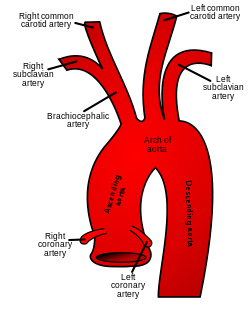 Plan of the branches. | |
 The thoracic aorta, viewed from the left side. | |
| Details | |
| Precursor | Dorsal aorta |
| Source | Ascending aorta |
| Branches | Thoracic aorta Abdominal aorta |
| Identifiers | |
| Latin | aorta descendens, pars descendens aortae |
| TA98 | A12.2.10.001 |
| TA2 | 4185 |
| FMA | 3784 |
| Anatomical terminology | |
In human anatomy, the descending aorta is part of the aorta, the largest artery in the body. The descending aorta begins at the aortic arch and runs down through the chest and abdomen. The descending aorta anatomically consists of two portions or segments, the thoracic and the abdominal aorta, in correspondence with the two great cavities of the trunk in which it is situated. Within the abdomen, the descending aorta branches into the two common iliac arteries which serve the pelvis and eventually legs.
The ductus arteriosus connects to the junction between the pulmonary artery and the descending aorta in foetal life. This artery later regresses as the ligamentum arteriosum.[1][2]
YouTube Encyclopedic
-
1/3Views:154 2294 80946 085
-
Thoracic (Descending) Aorta: Anatomy & Branches | Kenhub
-
Descending Thoracic Aorta Branches | Anatomy Made Easy
-
Clinical Anatomy - The Aorta, sections and branches (coarctation, dissection and aneurysm)
Transcription
Hello everyone! This is Matt from Kenhub, and this tutorial will describe the thoracic aorta and its branches. The aorta seen here, highlighted in green, is the largest blood vessel in the body, and almost all arteries stem from this vessel or from one of its branches. Our focus in this video is a section of the aorta called the thoracic aorta or descending aorta and its branches. You can see the clear view of it here without any organs in the way. The terms thoracic aorta and descending aorta are used interchangeably, and they're both perfectly acceptable. Here's an overview of the aorta so that you can find your bearings when we discuss the specifics of the thoracic aorta. After starting at the aortic valve, the ascending aorta winds towards the head, becomes the aortic arch or the transverse aorta as it makes a rainbow over the superior aspect of the heart, and then moves in an inferior direction through the chest and abdomen. At the left subclavian artery, it becomes the thoracic aorta. After the thoracic aorta, comes the abdominal aorta and follows, then, the thoracoabdominal aorta which ends in its bifurcation into the left and right common iliac arteries. Let's now focus on the thoracic aorta. It begins at the level of the fourth thoracic vertebra and descends on the left side of the thoracic vertebrae from the fifth thoracic vertebra to the 12th thoracic vertebra. Running behind the base of the left lung and the pericardium, it enters the abdomen via the aortic hiatus of the diaphragm when it reaches the T12 vertebra. The branches of the thoracic aorta include the bronchial arteries, the pericardial arteries, the superior phrenic arteries, the esophageal arteries, the posterior intercostals arteries, and the subcostal arteries. Once again, those branches and the parts they supply: the bronchial which supplies the lungs, the pericardial which supplies the dorsal portion of the pericardium, the superior phrenic supporting the diaphragm and the adrenal glands, the esophageal supplying the (you guessed it!) the esophagus. The posterior intercostal, which you see here in green, supply the intercostal spaces and the subcostal, the flat abdominal wall muscles. This video is more fun than reading a textbook, right? If you want more videos, interactive quizzes, articles, and an atlas of human anatomy, click on the "Take me to Kenhub" button. It is time to say goodbye to your old textbooks and say hello to your new anatomy learning partner, Kenhub! See you there! https://www.kenhub.com
See also
References
![]() This article incorporates text in the public domain from page 598 of the 20th edition of Gray's Anatomy (1918)
This article incorporates text in the public domain from page 598 of the 20th edition of Gray's Anatomy (1918)
- ^ Rubin, Raphael; Strayer, David S., eds. (2008). Rubin's Pathology: Clinicopathologic Foundations of Medicine (5th ed.). Philadelphia: Lippincott Williams & Wilkins. p. 442. ISBN 9780781795166.
- ^ Naidich, David P.; Webb, W. Richard; et al., eds. (2007). Computed Tomography and Magnetic Resonance of the Thorax (4th ed.). Philadelphia: Lippincott Williams & Wilkins. p. 100. ISBN 9780781757652.
External links
- Anatomy figure: 19:04-03 at Human Anatomy Online, SUNY Downstate Medical Center – "Left side of the mediastinum."
This cardiovascular system article is a stub. You can help Wikipedia by expanding it. |