


Rootletin also known as ciliary rootlet coiled-coil protein (CROCC) is a protein that in humans is encoded by the CROCC gene.[5][6][7] Rootletin is a component of the ciliary rootlet, and, together with CEP68 and CEP250, is required for centrosome cohesion.[8]
Rootletin is an important protein in the ciliary rootlet, particular for the structure and can be considered an important protein in mitosis as it is a centrosome linker.
YouTube Encyclopedic
-
1/1Views:430 042
-
Cytoskeleton Microtubules | Cell Biology
Transcription
Function
This protein forms part of the ciliary rootlet structure. It also helps to contribute to the centrosome cohesion before mitosis.[9] Expression of rooletin leads to the formation of fibrous protein.
Structure
This protein is part of the structure of a ciliary rootlet. This cytoskeletal-like structure starts from the basal body at one end of the cilium and extends towards nucleus. Its molecular structure consists of a globular head domain and a tail domain made up of coiled-coil structures.[5]
Protein interactions
A large coiled-coil protein, C-Nap1, is a docking site for the fibrous tether to proximal ends of centrioles which Rootletin physically interacts with. Furthermore, Rootletin is phosphorylated by Nek2 kinase.[10]
References
- ^ a b c GRCh38: Ensembl release 89: ENSG00000058453 – Ensembl, May 2017
- ^ a b c GRCm38: Ensembl release 89: ENSMUSG00000040860 – Ensembl, May 2017
- ^ "Human PubMed Reference:". National Center for Biotechnology Information, U.S. National Library of Medicine.
- ^ "Mouse PubMed Reference:". National Center for Biotechnology Information, U.S. National Library of Medicine.
- ^ a b Yang J, Liu X, Yue G, Adamian M, Bulgakov O, Li T (Nov 2002). "Rootletin, a novel coiled-coil protein, is a structural component of the ciliary rootlet". J Cell Biol. 159 (3): 431–40. doi:10.1083/jcb.200207153. PMC 2173070. PMID 12427867.
- ^ McClintock TS, Glasser CE, Bose SC, Bergman DA (Jan 2008). "Tissue expression patterns identify mouse cilia genes". Physiol Genomics. 32 (2): 198–206. doi:10.1152/physiolgenomics.00128.2007. PMID 17971504.
- ^ "Entrez Gene: CROCC ciliary rootlet coiled-coil, rootletin".
- ^ Graser S, Stierhof YD, Nigg EA (December 2007). "Cep68 and Cep215 (Cdk5rap2) are required for centrosome cohesion". J. Cell Sci. 120 (Pt 24): 4321–31. doi:10.1242/jcs.020248. PMID 18042621.
- ^ Bahe S, Stierhof YD, Wilkinson CJ, Leiss F, Nigg EA (October 2005). "Rootletin forms centriole-associated filaments and functions in centrosome cohesion". J. Cell Biol. 171 (1): 27–33. doi:10.1083/jcb.200504107. PMC 2171225. PMID 16203858.
- ^ Lim HH, Zhang T, Surana U (July 2009). "Regulation of centrosome separation in yeast and vertebrates: common threads". Trends Cell Biol. 19 (7): 325–33. doi:10.1016/j.tcb.2009.03.008. PMID 19576775.
Further reading
- Ching YP, Chan SF, Jeang KT, Jin DY (2006). "The retroviral oncoprotein Tax targets the coiled-coil centrosomal protein TAX1BP2 to induce centrosome overduplication". Nat. Cell Biol. 8 (7): 717–24. doi:10.1038/ncb1432. hdl:10722/54244. PMID 16767081. S2CID 10140583.
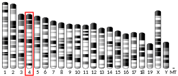
- Gregory SG, Barlow KF, McLay KE, et al. (2006). "The DNA sequence and biological annotation of human chromosome 1". Nature. 441 (7091): 315–21. Bibcode:2006Natur.441..315G. doi:10.1038/nature04727. PMID 16710414.
- Andersen JS, Wilkinson CJ, Mayor T, et al. (2003). "Proteomic characterization of the human centrosome by protein correlation profiling". Nature. 426 (6966): 570–4. Bibcode:2003Natur.426..570A. doi:10.1038/nature02166. PMID 14654843. S2CID 4427303.
- Behrends U, Schneider I, Rössler S, et al. (2003). "Novel tumor antigens identified by autologous antibody screening of childhood medulloblastoma cDNA libraries". Int. J. Cancer. 106 (2): 244–51. doi:10.1002/ijc.11208. PMID 12800201.
- Strausberg RL, Feingold EA, Grouse LH, et al. (2003). "Generation and initial analysis of more than 15,000 full-length human and mouse cDNA sequences". Proc. Natl. Acad. Sci. U.S.A. 99 (26): 16899–903. Bibcode:2002PNAS...9916899M. doi:10.1073/pnas.242603899. PMC 139241. PMID 12477932.
- Seki N, Ohira M, Nagase T, et al. (1998). "Characterization of cDNA clones in size-fractionated cDNA libraries from human brain". DNA Res. 4 (5): 345–9. doi:10.1093/dnares/4.5.345. PMID 9455484.