
| Brachioradialis muscle | |
|---|---|
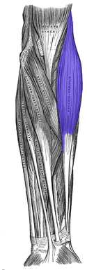 Anterior view of muscles of the left forearm with brachioradialis shown in blue | |
 Cross-section through the middle of the forearm. Brachioradialis labeled at center left, sixth from the top. | |
| Details | |
| Origin | Lateral supracondylar ridge of the humerus and the orbicular ligament of the radius |
| Insertion | Distal radius (radial styloid process) |
| Artery | Radial recurrent artery |
| Nerve | Radial nerve (C5-C8 & T1) |
| Actions | Flexion of elbow, supination and pronation of the radioulnar joint to 90° |
| Identifiers | |
| Latin | musculus brachioradialis |
| TA98 | A04.6.02.039 |
| TA2 | 2496 |
| FMA | 38485 |
| Anatomical terms of muscle | |
The brachioradialis is a muscle of the forearm that flexes the forearm at the elbow.[1][2] It is also capable of both pronation and supination, depending on the position of the forearm.[2] It is attached to the distal styloid process of the radius by way of the brachioradialis tendon, and to the lateral supracondylar ridge of the humerus.
YouTube Encyclopedic
-
1/5Views:39 88351 3074 5679 3936 945
-
Anatomy Of The Brachioradialis Muscle - Everything You Need To Know - Dr. Nabil Ebraheim
-
Coracobrachialis Brachialis Brachioradialis - Everything You Need To Know - Dr. Nabil Ebraheim
-
Two Minutes of Anatomy: Brachioradialis Muscle
-
Radial muscles of the forearm (preview) - Human Anatomy | Kenhub
-
How To Treat Trigger Points - Brachioradialis
Transcription
Structure
The brachioradialis is a superficial, fusiform muscle on the lateral side of the forearm. It originates proximally on the lateral supracondylar ridge of the humerus. It inserts distally on the radius, at the base of its styloid process.[3] Near the elbow, it forms the lateral limit of the cubital fossa, or elbow pit.[4]
Nerve supply
Despite the bulk of the muscle body being visible from the anterior aspect of the forearm, the brachioradialis is a posterior compartment muscle and consequently is innervated by the radial nerve. Of the muscles that receive innervation from the radial nerve, it is one of only four that receive input directly from the radial nerve. The other three are the triceps, anconeus, and extensor carpi radialis longus. (All other posterior compartment muscles that receive radial innervation are supplied by the deep branch of the radial nerve.)[citation needed]
Function
The brachioradialis flexes the forearm at the elbow.[1][2] When the forearm is pronated, the brachioradialis tends to supinate as it flexes.[2] In a supinated position, it tends to pronate as it flexes.[2] This also assists the biceps brachii.[2]
The brachioradialis is a stronger elbow flexor when the forearm is in a midposition between supination and pronation at the radioulnar joint. When pronated, the brachioradialis is more active during elbow flexion since the biceps brachii is in a mechanical disadvantage.
With the insertion of the muscle so far from the fulcrum of the elbow, the brachioradialis does not generate as much joint torque as the brachialis or the biceps. It is effective mainly when those muscles have already partially flexed at the elbow. The brachioradialis flexes the forearm at the elbow, especially when quick movement is required and when a weight is lifted during slow flexion of the forearm.
The muscle is used to stabilize the elbow during rapid flexion and extension while in a midposition, such as in hammering. The brachioradialis is synergistic with the brachialis and biceps brachii; the triceps brachii and anconeus are antagonistic.[5][6]
Additional images
-
 Left humerus. Anterior view.
 -
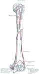
 Bones of left forearm. Anterior aspect.
 -
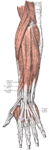
 Front of the left forearm. Superficial muscles.
 -
 Posterior surface of the forearm. Superficial muscles.
 -
 The radial artery.
 -
 The radial and ulnar arteries.
 -
 Ulnar and radial arteries. Deep view.
 -
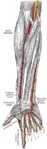
 Nerves of the left upper extremity.
 -
 Brachioradialis


References
- ^ a b Johnson, G. R. (2014-01-01), Revell, P. A. (ed.), "1 - Developments in joint replacement technology*Note: This chapter is an updated version of Chapter 1 from the first edition of Joint replacement technology edited by P. A. Revell and published by Woodhead Publishing, 2008.*", Joint Replacement Technology, Woodhead Publishing, pp. 3–30, doi:10.1533/9780857098474.1.3, ISBN 978-0-85709-841-2, retrieved 2020-12-12
- ^ a b c d e f Day, Judd S. (2016-01-01), Kurtz, Steven M. (ed.), "12 - The Clinical Performance of UHMWPE in Elbow Replacements", UHMWPE Biomaterials Handbook (Third Edition), Oxford: William Andrew Publishing, pp. 179–196, doi:10.1016/b978-0-323-35401-1.00012-0, ISBN 978-0-323-35401-1, retrieved 2020-12-12
- ^ Freehafer, Alvin A.; Hunter Peckham, P.; Keith, Michael W.; Mendelson, Laurel S. (1988-01-01). "The brachioradialis: Anatomy, properties, and value for tendon transfer in the tetraplegic". The Journal of Hand Surgery. 13 (1): 99–104. doi:10.1016/0363-5023(88)90210-9. ISSN 0363-5023. PMID 3351238.
- ^ Moore, KL.; Dalley, AF.; Agur, AM. (2013). Clinically Oriented Anatomy, 7th ed. Lippincott Williams & Wilkins. pp. 751–752. ISBN 978-1-4511-8447-1.
- ^ Bowden, Bradley S. Bowden, Joan M. An Illustrated Atlas of Skeletal Muscles. 2nd ed. 2002
- ^ Saladin, Kenneth S. Anatomy & Physiology. 4th ed. 2007
External links
- Illustration: upper-body/brachialis from The Department of Radiology at the University of Washington
- Anatomy figure: 07:01-09 at Human Anatomy Online, SUNY Downstate Medical Center - "Transverse section through the left arm just proximal to the elbow."
- Anatomy figure: 07:03-07 at Human Anatomy Online, SUNY Downstate Medical Center - "Superficial muscles of the anterior (flexor) compartment of the left forearm."
- Anatomy figure: 09:02-02 at Human Anatomy Online, SUNY Downstate Medical Center - "Superficial muscles of the posterior (extensor) compartment of the left forearm."
- lesson5musofpostforearm at The Anatomy Lesson by Wesley Norman (Georgetown University)