
| Brachialis | |
|---|---|
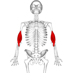
 Deep muscles of the chest and front of the arm, with the boundaries of the axilla. (Brachialis visible at bottom right.) | |
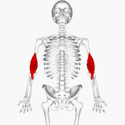
 Position of brachialis (shown in red). | |
| Details | |
| Origin | anterior surface of the humerus, particularly the distal half of this bone |
| Insertion | coronoid process and the tuberosity of the ulna |
| Artery | radial recurrent artery, brachial artery |
| Nerve | musculocutaneous nerve (C5-C7) and radial nerve (C5, C6) |
| Actions | flexion at elbow joint |
| Identifiers | |
| Latin | musculus brachialis |
| TA98 | A04.6.02.018 |
| TA2 | 2469 |
| FMA | 37667 |
| Anatomical terms of muscle | |
The brachialis (brachialis anticus) is a muscle in the upper arm that flexes the elbow. It lies beneath the biceps brachii, and makes up part of the floor of the region known as the cubital fossa (elbow pit). It originates from the anterior aspect of the distal humerus;[1] it inserts onto the tuberosity of the ulna. It is innervated by the musculocutaneous nerve,[2] and commonly also receives additional innervation from the radial nerve.[3] The brachialis is the prime mover of elbow flexion generating about 50% more power than the biceps.[dubious ][1]
YouTube Encyclopedic
-
1/5Views:155 6725 64968 18953 63021 116
-
Brachialis Muscle Anatomy Overview - Human Anatomy | Kenhub
-
Brachialis Muscle | Upper Limb
-
Brachialis Muscle Anatomy: Origin, Insertion, Workout Exercise, Action
-
Brachialis | Muscle Anatomy
-
Brachialis | Muscle Anatomy
Transcription
Hi, everyone. This is Matt from Kenhub! And in this tutorial, we will discuss the origin, insertion, innervation, and function of the brachialis muscle. The brachialis is a long, strong muscle of the upper arm. For the most part, the brachialis is buried under the biceps brachii and is therefore not easy to palpate from the surface. The muscle does have superficial parts found at its lateral border and distally. Even though it is located deep in the upper arm, the brachialis muscle still contributes indirectly to the surface anatomy as its large belly makes the biceps brachii look much larger on the surface than it actually is. You know how the old saying goes, “Behind every great biceps brachii is a great brachialis,” or something like that. The brachialis originates at the distal half of the anterior side of the humerus. In addition, the origin tendon attaches to the medial and lateral intermuscular septa of the arm, two dividing membranes separating the flexor from the extensor muscles. Distally, the muscle inserts at the tuberosity of the ulna, where its fibers are also connected to the joint capsule. The nerve supply for the brachialis comes from the musculocutaneous nerve. However, in 70% to 80% of people, the muscle has double innervation with the radial nerve. The brachialis is the strongest flexor of the elbow joint, stronger even than the biceps brachii because it is closer to the joint access, and furthermore, only stretches over one joint. A small contraction of the muscle consequently leads to a larger flexion in the elbow. Another function of the brachialis is helping with maintenance of tension found on the joint capsule, and as a result, working to prevent damages to the capsule during hyper extension. This video is more fun than reading a text book, right? If you want more videos, interactive quizzes, articles, and an atlas of human anatomy, click on the “Take me to Kenhub” button. It is time to say goodbye to your old textbooks, and say hello to your new anatomy learning partner, Kenhub. See you there! https://kenhub.com
Structure
Origin
The brachialis originates from the anterior surface of the distal half of the humerus,[1] near the insertion of the deltoid muscle, which it embraces by two angular processes. Its origin extends below to within 2.5 cm of the margin of the articular surface of the humerus at the elbow joint.[2]
Insertion
Its fibers converge to a thick tendon which is inserted into the tuberosity of the ulna,[2] and the rough depression on the anterior surface of the coronoid process of the ulna.[citation needed]
Innervation
The brachialis muscle is innervated by the musculocutaneous nerve, which runs on its superficial surface, between it and the biceps brachii.[2] However, in 70-80% of people, the muscle has double innervation with the radial nerve (C5-T1). The divide between the two innervations is at the insertion of the deltoid.[3]
Blood supply
The brachialis is supplied by muscular branches of the brachial artery and by the recurrent radial artery.[4]
Variation
The muscle is occasionally doubled; additional muscle slips to the supinator, pronator teres, biceps brachii, lacertus fibrosus, or radius are more rarely found.[citation needed]
Function
The brachialis flexes the arm at the elbow joint.[2] Unlike the biceps, the brachialis does not insert on the radius, and does not participate in pronation and supination of the forearm.[2]
History
Etymology
The brachialis muscle[5] In classical Latin bracchialis means of or belonging to the arm,[6] and is derived from classical Latin bracchium,"arm".[6] The expression musculus brachialis is used in the current official anatomic nomenco Terminologia Anatomica.[7]
Additional images
-
 Position of brachialis (shown in red). Animation.
 -
 Still image.
 -
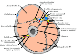 Horizontal section through the middle of upper arm. (Brachialis labeled at center left.)
 -
 Muscles of forearm, including insertion of brachialis tendon. Cross section. (Brachialis labeled at bottom left.)
 -
 Left humerus. Anterior view.
 -
 Bones of left forearm. Anterior aspect.
 -
 Nerves of the left upper extremity.
 -
 Brachialis muscle (labeled in green text)


See also
References
![]() This article incorporates text in the public domain from page 444 of the 20th edition of Gray's Anatomy (1918)
This article incorporates text in the public domain from page 444 of the 20th edition of Gray's Anatomy (1918)
- ^ a b c Saladin, Kenneth S, Stephen J. Sullivan, and Christina A. Gan. Anatomy & Physiology: The Unity of Form and Function. 2015. Print.
- ^ a b c d e f Drake, Richard L.; Vogl, Wayne; Tibbitts, Adam W.M. Mitchell; illustrations by Richard; Richardson, Paul (2005). Gray's anatomy for students. Philadelphia: Elsevier/Churchill Livingstone. p. 662,672. ISBN 978-0-8089-2306-0.
- ^ a b "Brachialis Muscle." Kenhub. Kenhub, Aug. 2001
- ^ "Brachialis." UW Department of Radiology. University of Washington, Nov. 2005
- ^ Di J.H. (Ed.) (1997).Stedman’s concise me10b">Triepel, H. (1910). Die anatomischen Namen. Ihre Ableitung und Aussprache. Mit eitte Auflage). Wiesbaden: Verlag J.F. Bergmann.
- ^ a b Lewis, C.T. & Short, C. (1879). A Latin dictionary founded on Andrews' edition of Freund's Latin dictionary. Oxford: Clarendon Press.
- ^ Federative Committee on Anatomical Terminology (FCAT) (1998). Terminologia Anatomica. Stuttgart: Thieme
External links
- Illustration: brachialis from The Department of Radiology at the University of Washington