
| Adductor magnus muscle | |
|---|---|
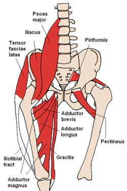 The adductor magnus and nearby muscles | |
 Structures surrounding right hip-joint (adductor magnus at upper right) | |
| Details | |
| Origin | Pubis, tuberosity of the ischium |
| Insertion | Linea aspera and adductor tubercle of femur |
| Artery | Deep femoral artery |
| Nerve | Posterior branch of obturator nerve (adductor) and sciatic nerve (hamstring) |
| Actions | Adduction of hip (both portions) flexion of hip (adductor portion) extension of hip (hamstring portion) |
| Identifiers | |
| Latin | musculus adductor magnus |
| TA98 | A04.7.02.028 |
| TA2 | 2630 |
| FMA | 22443 |
| Anatomical terms of muscle | |
The adductor magnus is a large triangular muscle, situated on the medial side of the thigh.
It consists of two parts. The portion which arises from the ischiopubic ramus (a small part of the inferior ramus of the pubis, and the inferior ramus of the ischium) is called the pubofemoral portion, adductor portion, or adductor minimus, and the portion arising from the tuberosity of the ischium is called the ischiocondylar portion, extensor portion, or "hamstring portion". Due to its common embryonic origin, innervation, and action the ischiocondylar portion (or hamstring portion) is often considered part of the hamstring group of muscles. The ischiocondylar portion of the adductor magnus is considered a muscle of the posterior compartment of the thigh while the pubofemoral portion of the adductor magnus is considered a muscle of the medial compartment.
YouTube Encyclopedic
-
1/5Views:83 4009 90010 4602 69217 201
-
Adductor Magnus Muscle - Function & Origins - Human Anatomy | Kenhub
-
Anatomy Of The Adductor Magnus Muscle - Everything You Need To Know - Dr. Nabil Ebraheim
-
Functions of the adductor magnus muscle (preview) - Human 3D Anatomy | Kenhub
-
The Adductor Magnus Muscle
-
Adductor Magnus 3D Animation 4k
Transcription
Hello again, everyone. It’s Matt from Kenhub. And in this tutorial, we will discuss the origin, insertion, innervation, and function of the adductor magnus. The adductors of the hip are part of the inner hip musculature and range from the lower pelvic bone to the femur and knee region. The hip adductors shape the surface anatomy of the medial thigh. This focus of this tutorial will be the adductor magnus. The adductor magnus muscle is one of the biggest muscles of the human body. It originates at the inferior pubic ramus, the ischial ramus, and the ischial tuberosity… And inserts both at the linea aspera and the adductor tubercle of the medial epicondyle. The innervation is mainly supplied by the obturator nerve which arises from the lumbar plexus and reaches the adductors through the obturator canal. The superficial part of the adductor magnus is supplied by the tibial nerve. As the name suggests, the main function of the hip adductors is the adduction of the hip joint. The adductor magnus additionally supports outward rotation, inward rotation, flexion, and extension. Overall, the hip adductors play an important role in balancing the pelvis during standing and walking. This video is more fun than reading a textbook, right? If you want more videos, interactive quizzes, articles, and an atlas of human anatomy, click on the “Take me to Kenhub” button. It is time to say goodbye to your old textbooks and say hello to your new anatomy learning partner, Kenhub! See you there! https://www.kenhub.com
Structure
Pubofemoral (adductor) portion
Those fibers which arise from the ramus of the pubis are short, horizontal in direction, and are inserted into the rough line of the femur leading from the greater trochanter to the linea aspera, medial to the gluteus maximus.
Those fibers from the ramus of the ischium are directed downward and laterally with different degrees of obliquity, to be inserted, by means of a broad aponeurosis, into the linea aspera and the upper part of its medial prolongation below.
Ischiocondylar (hamstring) portion
The medial portion of the muscle, composed principally of the fibers arising from the tuberosity of the ischium, forms a thick fleshy mass consisting of coarse bundles which descend almost vertically, and end about the lower third of the thigh in a rounded tendon which is inserted into the adductor tubercle on the medial condyle of the femur, and is connected by a fibrous expansion to the line leading upward from the tubercle to the linea aspera.
Relations
By its anterior surface the adductor magnus is in relation with the pectineus, adductor brevis, adductor longus, femoral artery and vein, profunda artery and vein, with their branches, and with the posterior branches of the obturator artery, obturator vein and obturator nerve.
By its posterior surface with the semitendinosus, semimembranosus, biceps, and gluteus maximus muscle.
By its inner border with the gracilis and sartorius.
By its upper border with the obturator externus, and quadratus femoris.[1]
Nerve supply
It is a composite muscle as the adductor and hamstring portions of the muscle are innervated by two different nerves. The adductor portion is innervated by the posterior division of the obturator nerve while the hamstring portion is innervated by the sciatic nerve.[2][3]
Osseoaponeurotic openings
At the insertion of the muscle, there is a series of osseoaponeurotic openings, formed by tendinous arches attached to the bone. The upper four openings are small, and give passage to the perforating branches of the profunda femoris artery. The lowest (often referred to as the adductor hiatus) is large, and transmits the femoral vessels to the popliteal fossa.
Variation
The upper, lateral part of the adductor magnus is an incompletely separated division often considered a separate muscle — the adductor minimus.[4] These two muscles are frequently separated by a branch of the superior perforating branch of the profunda femoris artery.[5]
Function
The adductor magnus is a powerful adductor of the thigh, made especially active when the legs are moved from a wide spread position to one in which the legs parallel each other. The part attached to the linea aspera acts as a lateral rotator. The part which reaches the medial epicondyle acts as a medial rotator when the leg is rotated outwards and flexed, and also acts to extend the hip joint.[4]
Other animals
In other tetrapods, the adductor magnus crosses the knee joint and inserts into the tibia. In humans, the distal part of the tendon detaches and becomes the medial collateral ligament of the knee. Because of this, the medial collateral ligament of the knee in humans may contain a few muscle fibres as an atavistic variation.[6]
Additional images
-
 Muscles of the iliac and anterior femoral regions.
 -
 Cross-section through the middle of the thigh.
 -
 Deep muscles of the medial femoral region.
 -
 The arteries of the gluteal and posterior femoral regions.
 -
 Adductor magnus muscle
 -
 Adductor magnus muscle
 -
 Muscles of Thigh. Anterior views.
 -
 Muscles of Thigh. Anterior views.


See also
References
![]() This article incorporates text in the public domain from page 473 of the 20th edition of Gray's Anatomy (1918)
This article incorporates text in the public domain from page 473 of the 20th edition of Gray's Anatomy (1918)
- ^ Wilson, Erasmus (1851) The anatomist's vade mecum: a system of human anatomy, p 261
- ^ MedicalMnemonics.com: 255
- ^ "Adductor Magnus". Department of Radiology, University of Washington. Archived from the original on 28 August 2010. Retrieved 2010-08-16.
- ^ a b Platzer, Werner (2004), " Color Atlas of Human Anatomy, Vol. 1, Locomotor System, Thieme, 5th ed, p 242
- ^ Bergman, Ronald A.; Afifi, Adel K.; Miyauchi, Ryosuke (2010), Adductor Minimus (Henle, Günther), Anatomy Atlases
- ^ Norman Eizenberg et al., General Anatomy: Principles and Applications (2008), p 53.
External links
- Anatomy photo:14:st-0401 at the SUNY Downstate Medical Center
- PTCentral