
| Abductor pollicis longus muscle | |
|---|---|
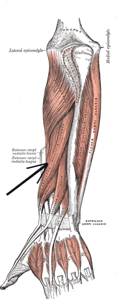 Deep muscles of posterior surface of the forearm | |
| Details | |
| Origin | ulna, radius, Interosseous membrane |
| Insertion | Base of 1st metacarpal |
| Artery | Posterior interosseous artery |
| Nerve | Radial nerve C8, T1 |
| Actions | abduction, extension of thumb |
| Antagonist | Adductor pollicis muscle |
| Identifiers | |
| Latin | musculus abductor pollicis longus |
| TA98 | A04.6.02.049 |
| TA2 | 2517 |
| FMA | 38515 |
| Anatomical terms of muscle | |
In human anatomy, the abductor pollicis longus (APL) is one of the extrinsic muscles of the hand. Its major function is to abduct the thumb at the wrist. Its tendon forms the anterior border of the anatomical snuffbox.
YouTube Encyclopedic
-
1/5Views:63 1972 5361 1651 98038 087
-
Abductor Pollicis Longus Muscle Overview - Human Anatomy | Kenhub
-
Abductor Pollicis Longus | Muscle Anatomy | Doctor Speaks
-
Abductor Pollicis Longus Musclepath, Origin, Insertion 3D Animation
-
Abductor Pollicis Longus Function: Thumb Abduction (3D Animation)
-
Abductor Pollicis Longus - Everything You Need To Know - Dr. Nabil Ebraheim
Transcription
Structure
The abductor pollicis longus lies immediately below the supinator and is sometimes united with it. It arises from the lateral part of the dorsal surface of the body of the ulna,[1] below the insertion of the anconeus, from the interosseous membrane, and from the middle third of the dorsal surface of the body of the radius.[2]
Passing obliquely downward and lateralward, it ends in a tendon, which runs through a groove on the lateral side of the lower end of the radius, accompanied by the tendon of the extensor pollicis brevis.[2]
The insertion is divided into a distal, superficial part and a proximal, deep part. The superficial part is inserted with one or more tendons into the radial side of the base of the first metacarpal bone, and the deep part is variably inserted into the trapezium, the joint capsule and its ligaments, and into the belly of abductor pollicis brevis (APB) or opponens pollicis.[3]
Innervation
The abductor pollicis longus muscle is innervated by the posterior interosseous nerve, which is a continuation of the deep branch of the radial nerve after it passes through the supinator muscle. Abductor pollicis longus lies close to the radial nerve.[4] The posterior interosseous nerve is derived from spinal segments C7 & C8.[5]
Blood supply
Abductor pollicis longus is supplied by the posterior interosseous artery.[6]
Variation
An accessory abductor pollicis longus (AAPL) tendon is present in more than 80% of people, and a separate muscle belly is present in 20% of people. In one study, the accessory tendon was inserted into the trapezium (41%); proximally on the abductor pollicis brevis (22%) and opponens pollicis brevis (5%); had a double insertion on the trapezium and thenar muscles (15%); or the base of the first metacarpal (1%).[7] Up to seven tendons have been reported in rare cases.[8]
Multiple APL tendons can be regarded as a functional advantage since injured tendons can be compensated by the healthy ones.[9]
Function
The chief action of abductor pollicis longus is to abduct the thumb at the carpometacarpal joint, thereby moving the thumb anteriorly. It also assists in extending and rotating the thumb.[6]
By its continued action, it helps to abduct the wrist (radial deviation) and flex the hand.[6]
The APL insertion on the trapezium and the APB origin on the same bone is the only connection between the thumb's intrinsic and extrinsic muscles.[a] As the thumb is brought into action, these two muscles must coordinate to keep the trapezium stable in the carpus,[3] which is important for the proper functioning of the thumb (i.e. precision and power grip.)[10]
In other animals
The only primates to have an APL completely separated from the extensor pollicis brevis are modern humans and gibbons.[11] In gibbons, however, the APL originates proximally on the radius and ulna, whereas it originates in the middle part of these bones in crab-eating monkeys, bonobos, and humans. In all these primates, the muscle is inserted onto the base of the first metacarpal and sometimes onto the trapezium (siamangs and bonobos) and thumb sesamoids (crab-eating monkeys).[12]
In chimpanzees, the APL flexes the thumb rather than extends it like in modern humans. Compared to the wrists of chimpanzees, the human wrist is derived (compared to the Pan-Homo LCA) in having considerably longer muscle moment arms for a range of hand muscles. It is possible that these differences are due to the supinated position of the trapezium in humans which, in its turn, is a result of the expansion of the trapezoid on the side of the palm.[13]
A small, lens-shaped radial sesamoid embedded into the APL tendon is a primitive state found in all known Carnivora genera except in the red and giant pandas and the extinct Simocyon where it is hypertrophied (enlarged) into a sixth digit or a so-called "false thumb", a derived trait that first appeared in ursids.[14] The APL sesamoid is present in all non-human primates, but only in about half of gorillas, and normally absent in humans.[15]
Gallery
-
 Abductor pollicis longus muscle
 -
 Abductor pollicis longus muscle
 -
 Abductor pollicis longus muscle
 -
 Abductor pollicis longus muscle
 -
 Abductor pollicis longus muscle
 -
 Abductor pollicis longus muscle
 -
 Abductor pollicis longus muscle
 -
 Muscle of the hand. Posterior view.
See also
Notes
- ^ The extrinsic thumb muscles are those originating in the forearm: extensor pollicis longus, extensor pollicis brevis, flexor pollicis longus, and abductor pollicis longus. The intrinsic thumbs muscles originates in the hand: opponens pollicis, flexor pollicis brevis, adductor pollicis and abductor pollicis brevis.
References
- ^ Focks, Claudia; März, Ulrich (2008-01-01), Focks, Claudia (ed.), "Chapter 4 - Acupuncture Points of the Twelve Primary Channels", Atlas of Acupuncture, Edinburgh: Churchill Livingstone, pp. 79–462, doi:10.1016/b978-044310028-4.50007-2, ISBN 978-0-443-10028-4, retrieved 2020-10-22
- ^ a b Gray's Anatomy (1918), see infobox
- ^ a b van Oudenaarde & Oostendorp 1995
- ^ Bouche, P. (2013-01-01), Said, Gérard; Krarup, Christian (eds.), "Chapter 19 - Compression and entrapment neuropathies", Handbook of Clinical Neurology, Peripheral Nerve Disorders, Elsevier, 115: 311–366, doi:10.1016/b978-0-444-52902-2.00019-9, ISBN 9780444529022, PMID 23931789, retrieved 2020-10-22
- ^ "Abductor pollicis longus". GPnotebook. Retrieved 25 September 2016.
- ^ a b c "Abductor pollicis longus". PT Central. 1998. Archived from the original on 2012-02-04. Retrieved 25 September 2016.
- ^ Bravo, Barco & Bullón 2010, Results. For a dissection example see Fig. 3 in Hazani et al. 2008
- ^ Mehta et al. 2009, Discussion
- ^ Mehta et al. 2009, Conclusions
- ^ van Oudenaarde 1991, Introduction
- ^ Aversi-Ferreira et al. 2011, Results and Discussion
- ^ Michilsens et al. 2009
- ^ Tocheri et al. 2008, The evolution of the hominin hand as evidenced by the fossil record
- ^ Salesa et al. 2006
- ^ Le Minor 1994, Abstract
Sources
- Aversi-Ferreira, Tales Alexandre; Maior, Rafael Souto; Carneiro-e-Silva, Frederico O.; Aversi-Ferreira, Roqueline A. G. M. F.; Tavares, Maria Clotilde; Nishijo, Hisao; Tomaz, Carlos (2011). "Comparative Anatomical Analyses of the Forearm Muscles of Cebus libidinosus (Rylands et al. 2000): Manipulatory Behavior and Tool Use". PLOS ONE. 6 (7/e22165): e22165. Bibcode:2011PLoSO...622165A. doi:10.1371/journal.pone.0022165. PMC 3137621. PMID 21789230.
- Bravo, Elena; Barco, Raul; Bullón, Adrian (May 2010). "Anatomic Study of the Abductor Pollicis Longus: A Source for Grafting Material of the Hand". Clin Orthop Relat Res. 468 (5): 1305–1309. doi:10.1007/s11999-009-1059-4. PMC 2853646. PMID 19760470.
- Hazani, Ron; Engineer, Nitin J.; Cooney, Damon; Wilhelmi, Bradon J. (2008). "Anatomic Landmarks for the First Dorsal Compartment". ePlasty. 8 (e53): e53. PMC 2586286. PMID 19092992.
- Le Minor, JM (1994). "The sesamoid bone of musculus abductor pollicis longus (os radiale externum or prepollex) in primates". Acta Anat (Basel). 150 (3): 227–31. doi:10.1159/000147623. PMID 7817720.
- Mehta, Vandana; Arora, Jyoti; Suri, Rajesh Kumar; Rath, Gayatri (February 2009). "A Rare Quadruplicate Arrangement of Abductor Pollicis Longus Tendons — Anatomical and Clinical Relevance". Clinics. 64 (2): 153–155. doi:10.1590/S1807-59322009000200014. PMC 2666473. PMID 19219322.
- Michilsens, Fana; Vereecke, Evie E; D'Août, Kristiaan; Aerts, Peter (September 2009). "Functional anatomy of the gibbon forelimb: adaptations to a brachiating lifestyle". J. Anat. 215 (3): 335–354. doi:10.1111/j.1469-7580.2009.01109.x. PMC 2750765. PMID 19519640.
- van Oudenaarde, E (February 1991). "Structure and function of the abductor pollicis longus muscle". J. Anat. 174: 221–227. PMC 1256056. PMID 2032936.
- van Oudenaarde, E; Oostendorp, R A (June 1995). "Functional relationship between the abductor pollicis longus and abductor pollicis brevis muscles: an EMG analysis". J. Anat. 186 (Pt 3): 509–515. PMC 1167009. PMID 7559124.
- Salesa, Manuel J.; Antón, Mauricio; Peigné, Stéphane; Morales, Jorge (January 2006). "Evidence of a false thumb in a fossil carnivore clarifies the evolution of pandas". Proc Natl Acad Sci USA. 103 (2): 379–382. Bibcode:2006PNAS..103..379S. doi:10.1073/pnas.0504899102. PMC 1326154. PMID 16387860.
- Tocheri, Matthew W.; Orr, Caley M.; Jacofsky, Marc C.; Marzke, Mary W. (April 2008). "The evolutionary history of the hominin hand since the last common ancestor of Pan and Homo". J. Anat. 212 (4): 544–562. doi:10.1111/j.1469-7580.2008.00865.x. PMC 2409097. PMID 18380869.