
| Vitreous membrane | |
|---|---|
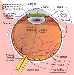 Schematic diagram of the human eye. Vitreous membrane is not labeled but it surrounds the vitreous, the dark mauve/purple area labeled 'Vitreous Humour'. | |
 The upper half of a sagittal section through the front of the eyeball. Vitreous membrane is not labeled but is visible. | |
| Details | |
| Identifiers | |
| Latin | membrana vitrea |
| TA98 | A15.2.06.012 |
| TA2 | 6813 |
| Anatomical terminology | |
The vitreous membrane (or hyaloid membrane or vitreous cortex) is a layer of collagen separating the vitreous humour from the rest of the eye. At least two parts have been identified anatomically. The posterior hyaloid membrane separates the rear of the vitreous from the retina. It is a false anatomical membrane.[1] The anterior hyaloid membrane separates the front of the vitreous from the lens.[2] Bernal et al. describe it "as a delicate structure in the form of a thin layer that runs from the pars plana to the posterior lens, where it shares its attachment with the posterior zonule via Weigert's ligament, also known as Egger's line".
YouTube Encyclopedic
-
1/3Views:31621 681683
-
Vitrectomy: PVR like something from Avatar.mpg
-
Vitrectomy: Peeling The Posterior Hyaloid VR1 Basic Techniqu
-
J Cannula Hooked the Capsule
Transcription
References
- ^ M P Snead, D R J Snead, A J Richards, J B Harrison, A V Poulson, A H C Morris, R M Sheard, J D Scott; Clinical, histological and ultrastructural studies of the posterior hyaloid membrane; "Eye", July 2002, Volume 16, Number 4, Pages 447-453.
- ^ Andres Bernal, Jean-Marie Parel, Fabrice Manns; Evidence for posterior zonular fiber attachment on the anterior hyaloid membrane; "Investigative Ophthalmology and Visual Science" 2006, 47, 4708-4713.
External links
This article about the eye is a stub. You can help Wikipedia by expanding it. |