
| Urogenital sinus | |
|---|---|
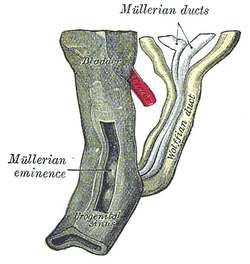 Urogenital sinus of female human embryo of eight and a half to nine weeks old. (Urogenital sinus labeled at bottom.) | |
 Stages in the development of the external sexual organs in the male and female. ("Opening of urogenital sinus" labeled in diagram D.) | |
| Details | |
| Carnegie stage | 15 |
| Precursor | Cloaca |
| Gives rise to | Urethra, bladder, vagina, vulval vestibule, Bartholin's glands, Skene's glands, prostate, bulbourethral glands |
| Identifiers | |
| Latin | sinus urogenitalis definitivus |
| TE | sinus_by_E5.7.3.1.0.0.1 E5.7.3.1.0.0.1 |
| Anatomical terminology | |
The urogenital sinus is a part of the human body only present in the development of the urinary and reproductive organs. It is the ventral part of the cloaca, formed after the cloaca separates from the anal canal during the fourth to seventh weeks of development.[1]
In males, the UG sinus is divided into three regions: upper, pelvic, and phallic. The upper part gives rise to the urinary bladder and the pelvic part gives rise to the prostatic and membranous parts of the urethra,[1] the prostate and the bulbourethral glands (Cowper's). The phallic portion gives rise to the spongy (bulbar) part of the urethra and the urethral glands (Littré's).
In females, the pelvic part of the UG sinus gives rise to the sinovaginal bulbs, structures that will eventually form the inferior two thirds of the vagina. This process begins when the lower tip of the paramesonephric ducts, the structures that will eventually form the uterus and vaginal fornices, come in contact with the UG sinus. Shortly afterwards, the sinovaginal bulbs form as two solid evaginations of the UG sinus. Cells in these bulbs divide to form a solid vaginal plate, which extends and then canalizes (hollows) to form the inferior portion of the vagina.[2] The female urogenital sinus also gives rise to the urethra, vestibule, Skene's glands and Bartholin's glands.
YouTube Encyclopedic
-
1/3Views:6 52912 473480 750
-
urogenital sinus
-
3D Embryology of Urinary Bladder & Urethra
-
Embryology | Development of Reproductive System
Transcription
Clinical significance
A urogenital sinus anomaly is also a rare birth defect in women where the urethra and vagina both open into a common channel.[3][4]
A persistent cloaca is a disorder where the rectum, vagina, and urinary tract meet and fuse, creating a cloaca, a single common channel.[5]
Other animals
In many mammals (excluding primates), the urogenital sinus refers to the sinus in which the openings to the female's urethra and vagina are found.[6] However, this term may also apply to some male mammals. The urogenital sinus of non-primates is homologous to the vulval vestibule of primates.
Additional images
-
 Enlarged view from the front of the left Wolffian body before the establishment of the distinction of sex.

See also
References
- ^ a b Sadler & Langman 2010, pp. 243–244.
- ^ Sadler & Langman 2010, pp. 253.
- ^ Callahan, Gerald N. (2009). Between XX and XY : intersexuality and the myth of two sexes. Chicago, Ill.: Chicago Review Press. p. 182. ISBN 978-1-56976-289-9. OCLC 436089205.
- ^ "Urogenital Sinus". Children's Hospital of Orange County. 2023-02-23. Retrieved 2023-10-07.
- ^ Jenkins D, Bitner-Glindzicz M, Thomasson L, et al. (2007), "Mutational analyses of UPIIIA, SHH, EFNB2 and HNF1β in persistent cloaca and associated kidney malformations", J Pediatr Urol, 3 (1): 2–9, doi:10.1016/j.jpurol.2006.03.002, PMC 1864944, PMID 17476318.
- ^ Schatten, Heide; Constantinescu, Gheorghe M. (2007-08-14). Comparative Reproductive Biology. John Wiley & Sons. ISBN 978-0-8138-1554-1.
Sources
- Sadler, T. W.; Langman, Jan (2010). Langman's Medical Embryology. Philadelphia: Lippincott William & Wilkins. ISBN 978-0-7817-9485-5. OCLC 1194422650.
External links
- "Pediatric Conditions - Abnormalities - Vaginal Anomalies: Urogenital Sinus". UrologyHealth.org. 2003-04-29. Archived from the original on 2004-02-18.
- "Fetal Pig Dissection". Medicine LibreTexts. 2020-10-15. Retrieved 2023-10-08.
- "Development of the reproductive system". Amboss.com. 2022-03-30.
- "Differentiation of the urogenital sinus in males". embryology.ch. 2007-09-27. Archived from the original on 2018-02-15.